Team:NEU CHINA/Engineering

Engineering Success
In the course of the project, the experimental results are analyzed timely. We would try the best to find out
drawbacks, and continuously improved the design and experiments to make our project much more better.
drawbacks, and continuously improved the design and experiments to make our project much more better.

Design
The PmrCAB two-component system is originally located in Salmonella. The Fe(III) sensitive domain were replaced by the receptor core domains of SARS-CoV-2, MERS-CoV, HCoV-229E respectively, to make the sensor protein PmrB sensitive to viral stimulation and respond to it as soon as possible. Therefore, the fluorescence signal expressed by the bacteria could be captured immediately and precisely.

Fig. 1 The recombinant PmrCAB detective system.
Fig. 1 The recombinant PmrCAB detective system.

Build
Three plasmids were constructed in pETDuet-1 as the vector of modified PmrCAB virus detection system. After transforming into E. coli BL21 (DE3), the first stage of detector was completed.

Fig. 2 A: The plasmid was constructed to detect SARS-CoV-2’s spike protein(BBa_K3611002). B: The plasmid was constructed to detect MERS-CoV’s spike protein(BBa_K3896007). C: The plasmid was constructed to detect HCoV-229E’s spike protein(BBa_K3896008).
Fig. 2 A: The plasmid was constructed to detect SARS-CoV-2’s spike protein(BBa_K3611002). B: The plasmid was constructed to detect MERS-CoV’s spike protein(BBa_K3896007). C: The plasmid was constructed to detect HCoV-229E’s spike protein(BBa_K3896008).
Test
To verify the validity of our bacteria, the characterization experiments are as follows.
When the bacteria grew to a suitable stage, IPTG was added to induce the expression of PmrB and PmrA. The appropriate amount of three kind of spike protein, corresponding to three types of coronavirus, and the sensor PmrB was stimulated to start a series of reactions to turn on the expression of downstream reporter gene by PmrC.
 Fig. 3 The fluorescence of engineered bacteria with ACE2 receptor
 Fig. 4 The fluorescence of engineered bacteria with DPP4 receptor.
 Fig. 5 The fluorescence of engineered bacteria with hAPN receptor.
When the bacteria grew to a suitable stage, IPTG was added to induce the expression of PmrB and PmrA. The appropriate amount of three kind of spike protein, corresponding to three types of coronavirus, and the sensor PmrB was stimulated to start a series of reactions to turn on the expression of downstream reporter gene by PmrC.
Fig. 3 The fluorescence of engineered bacteria with ACE2 receptor
Fig. 4 The fluorescence of engineered bacteria with DPP4 receptor.
Fig. 5 The fluorescence of engineered bacteria with hAPN receptor.
Analysis and learning
According to the result graph, it can be seen that the engineered bacteria receive the virus signal and finally successfully emit green fluorescence through a series of reactions. These results confirmed that our PmrCAB system is worked.
However, the concentration of spike protein added in the experiment is higher than that in the actual environment, thus solving the problem that virus titer might be extremely low in practical application. Thus, we focused on quorum sensing.
However, the concentration of spike protein added in the experiment is higher than that in the actual environment, thus solving the problem that virus titer might be extremely low in practical application. Thus, we focused on quorum sensing.
Design
We selected LuxI/LuxR quorum sensing system in E. coli.
When the engineered bacteria recognize the virus, PmrC initiates the expression of the downstream gene LuxI and LuxR. Catalyzed by LuxI, AHL starts to be synthesized continuously and part of it penetrates the cell membrane to the extracellular. When the concentration of extracellular AHL reaches a threshold, it re-enters the cell and combines with LuxR to form a LuxR-AHL co-complex, the co-complex can activate the corresponding promoter PLux_HS and express the reporter gene EGFP.
When the engineered bacteria recognize the virus, PmrC initiates the expression of the downstream gene LuxI and LuxR. Catalyzed by LuxI, AHL starts to be synthesized continuously and part of it penetrates the cell membrane to the extracellular. When the concentration of extracellular AHL reaches a threshold, it re-enters the cell and combines with LuxR to form a LuxR-AHL co-complex, the co-complex can activate the corresponding promoter PLux_HS and express the reporter gene EGFP.

Build
We added LuxI/LuxR quorum sensing system to the design and constructed the following two plasmids with pETDuet-1 and pACYCDuet-1 as the vectors, which were co-transformed into E. coli BL21 (DE3), and the vectors carried ampicillin and chloramphenicol resistance, respectively.
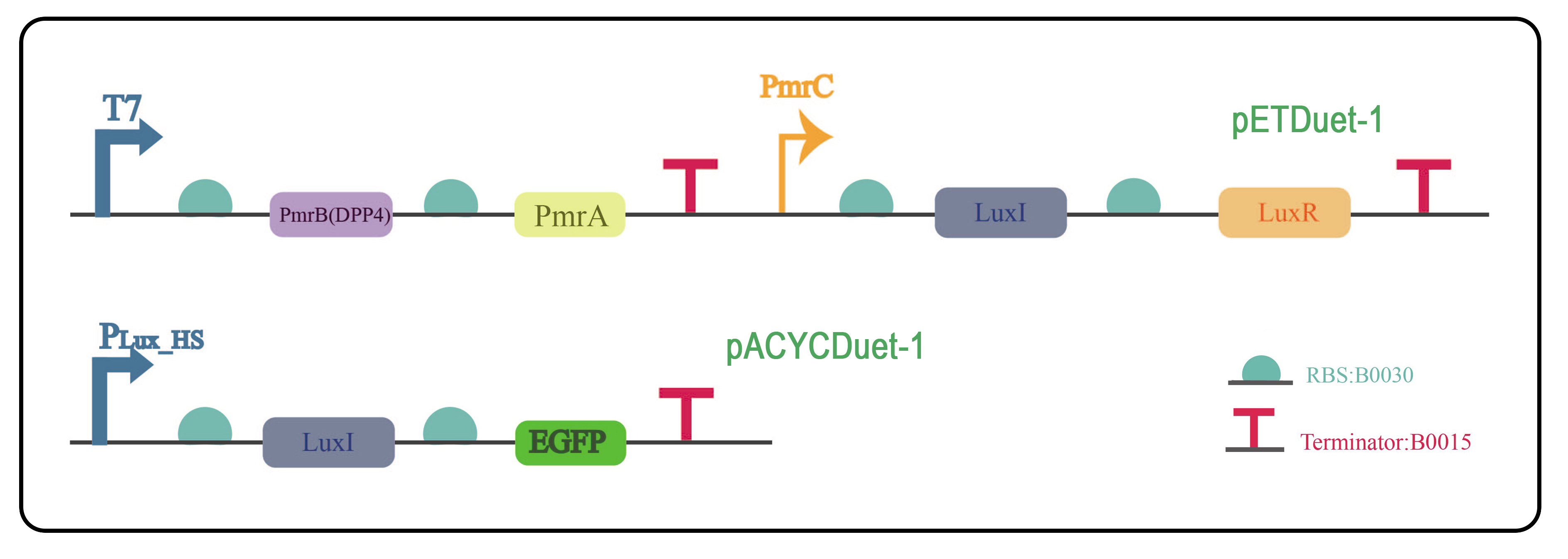
Fig. 6 The two plasmids were constructed with pETDuet-1 and pACYCDuet-1 as the vectors, and were co-transformed to E. coli BL21(DE3) to verify the quorum sensing system (BBa_K3896016 & BBa_K3896017).
Fig. 6 The two plasmids were constructed with pETDuet-1 and pACYCDuet-1 as the vectors, and were co-transformed to E. coli BL21(DE3) to verify the quorum sensing system (BBa_K3896016 & BBa_K3896017).
Test
To verify whether the quorum sensing system is effective. Our characterization experiments were as follows.
When the bacterial fluid OD reached certain conditions, IPTG was added to induce the expression of PmrB and PmrA, and then a small amount of Spike protein was added, and the fluorescence intensity and absorbance were measured every hour with an enzyme-labeled instrument, and the following are the results four hours after the addition of S protein.

Fig. 7 The fluorescence intensity of engineered bacteria without quorum sensing system and with quorum sensing.
When the bacterial fluid OD reached certain conditions, IPTG was added to induce the expression of PmrB and PmrA, and then a small amount of Spike protein was added, and the fluorescence intensity and absorbance were measured every hour with an enzyme-labeled instrument, and the following are the results four hours after the addition of S protein.
Fig. 7 The fluorescence intensity of engineered bacteria without quorum sensing system and with quorum sensing.
Analysis and learning
Based on the above results, we can see that the addition of quorum sensing can detect the results even if the amount of spike protein is reduced, which indicates that the LuxI/LuxR quorum sensing can improve the sensitivity of detection.
We noted that in this design, the extracellular concentration of AHL is the key element, which needs to reach a certain threshold and enter the cell for the quorum sensing to work. We learned from the literature that the threshold for AHL is typically 4-8 𝝁mol/L. We wanted to know at what concentration AHL in bacterial solution could enter the cell and bind to LuxR to form a complex in our actul experiment, so we performed the following design.
We noted that in this design, the extracellular concentration of AHL is the key element, which needs to reach a certain threshold and enter the cell for the quorum sensing to work. We learned from the literature that the threshold for AHL is typically 4-8 𝝁mol/L. We wanted to know at what concentration AHL in bacterial solution could enter the cell and bind to LuxR to form a complex in our actul experiment, so we performed the following design.
Design
We designed to add a range of concentrations of AHL that reached a threshold to enter the bacteria and bind to endogenous LuxR to form co-complex LuxR-AHL to activate the promoter and express the reporter gene EGFP.

Build
We successfully constructed and transformed the following plasmids into E. coli BL21 (DE3) using pACYCDuet-1 as a vector, which itself carries chloramphenicol resistance.

Fig. 8 The plasmid was constructed with pACYCDuet-1 as the vector to explore the concentration threshold of AHL.
Fig. 8 The plasmid was constructed with pACYCDuet-1 as the vector to explore the concentration threshold of AHL.
Test
We simulated the extracellular release of AHL from bacteria under natural conditions by artificially adding AHL. we set a concentration gradient of AHL (0 𝝁mol/L to 13 𝝁mol/L), waited for the bacteria to grow to a suitable period, added different concentrations of AHL, and AHL that reached the concentration threshold entered the engineered bacteria, combined with the endogenous LuxR to activate the PLux_HS promoter, and induced expression of downstream genes. After incubation for appropriate time, the fluorescence intensity and absorbance were measured. The results obtained are as follows.

Fig. 9 The fluorescence intensity of engineered bacteria under different concentrations of AHL.
Fig. 9 The fluorescence intensity of engineered bacteria under different concentrations of AHL.
Analysis and learning
Based on the above experimental results, we found that the fluorescence intensity would be enhanced with the increase of the concentration, and reached the peak at 9 𝝁mol/L. Continuing to increase the concentration, the experimental results no longer varied regularly.