Team:Korea HS/Experiments
KORHS
1st Lab Meeting
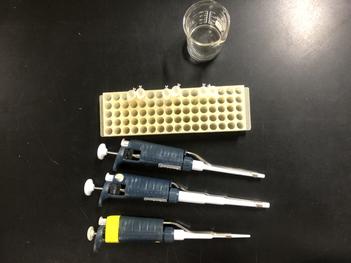
Micropipette information: MicroPipette (Gilson) was utilized for all experimental expenditures. It provided a convenient, cost-effective delivery method and required fewer chemicals to deliver a certain volume of fluid (maximum volume: 1mL = 1000μL / minimum volume: 0.2mL = 200μL - 1000/200/100/20/10/2P).
- 1. Place the tip of the pipette into disposable tips.
- 2. Press the pressure button (absorbs a certain volume of liquid).
- 3. Rotate rubber to set volume.
- 4. Press until first stop before uptaking liquid.
- 5. Press till 2nd stop to release all remaining chemicals (more safety and accuracy).
- 1. The solution should stay out of the pipet as contamination could occur.
- 2. Always hold the pipette vertically, tip down.
- 3. Every time the pipet enters the solution, the solution can change concentrations or be contaminated. Change the tip every time the chemical changes.
- 4. Press the release button to release the tip.
- 5. Place the tip on the left wall of the tube and let the liquid move down.
- 6. A lot of waste may be generated. Try minimizing waste for the environment.
- 7. When releasing liquid, always hold it straight, not slanted (refer to the law of gravity: all liquids flow downwards).

- 1. MRC5 (normal lung cell/fibroblast presenting slow cell division, thus displaying higher sensitivity and requiring careful handling)
- 2. A549 (lung cancer/carcinoma cell that proliferates much more rapidly)
- 3. Plastic cell culture plate (transparent for microscopy)
- 4. 6-well (with bigger individual wells) and/or 96-well plate (with smaller individual wells)
During cell culture experiments, two types of cell lines might ensue: adherent cell (growing on the bottom of medium) and suspension cell (not growing on the bottom of medium).

One method to cell detachment is by physical force, or scraping of cells. However, this method is not recommended as cells often burst and die off when scraped. Enzyme method, correcting limitations described hitherto, was utilized. Trypsin protease degrades intracellular proteins that bind cells together, thus inducing cleavage. It is a recommended protocol to keep a maximum of a 10-minute interval as cells will die when isolated - minimized interval with trypsin to avoid cell damage (i.e. suspended cells = apoptosis). 5-minute enzyme incubation time for trypsin to detach all cells was set. Cells in subculture were counted (cells/mL) to check if cells detach.
2nd Lab Meeting
In this study, three CPPs (derivative buforin IIb, analog MV1 and MV2) were tested. MV1 and MV2 were modified because smaller peptides enter cancer cells more easily.
Buforin IIb: RAGLQFPVGRLLRRLLRRLLR 21aa + charge (~3000Mw, 1000Mw = 1mg)MV1: RAGLQFPVGRLLRRLLR 17aa + charge (2524 Mw)
MV2: RAGLQFPVGRLLR 13aa + charge (1985 Mw)
Cell counter holds a lack of sensitivity. Thus, an MTT assay was performed in this study. MTT-yellow tetrazolium (substrate) is inserted in each well and mitochondrial reductase enzyme inside viable cells reduces substrate to purple-dyed formazan (reaction: yellow → purple). Purple-dyed formazan doesn’t absorb purple. Hence, the spectrometer measures absorbance.
The mathematically presented graph presents the x-axis (live cell number) and y-axis (absorbance at 570nm) with the following assumption: positive linear relationship = higher concentration = higher absorbance. That is, in reality, the graph measures the correlation between live cell number and mitochondria reductase activity. Overall, we aim to use a peptide with low cytotoxicity which leads to higher cell viability and minimizes side effects on normal cells.
- 1. 5 μL MTT solution insertion into 9 wells
- 2. 1-hour incubation for reaction completion
- 3. Supernatant (remaining media) removal
- 4. 100X or 100 μL solubilization solution application (cytolysis and formazan dye release)
- 5. 10 minutes incubation for reaction completion
- 6. 550-600nm spectrometer analysis

- ▪ A549 lung cancer cell in each well
- ▪ 1st nine: Cell media [control (healthy cancer cells)]
- ▪ 2nd nine: Dead cells [SDS → leads to degradation of proteins 20%]
- ▪ 3rd nine: Concentration-dependent (5, 10, 20, 40, 80, 100 micromoles)
- ▫ Assumption: The higher the concentration, the higher the cytotoxicity.
- ▫ Model: 5 μM first column MV2, second column MV1, third column: BIIB
- ▪ 4th nine: 10μM first column MV2, second column MV1, third column: BIIB
- ▫ Repeat the remaining nine(s) in a concentration-dependent manner.
- ▪ Each well = 100 μL total volume
- 1. DNA modeling, cloning, synthesis
- 2. Host cell insertion (decide if normal cell or bacteria will be used)
- 3. Protein translation
- 4. Protein extraction
- 5. Protein purification
However, CPP is a very small macromolecule of <30 amino acids. Thus, it renders purification difficult and cost-inefficient. Hence, without transfecting the bacterial host, the peptide synthesizer will bio-synthesize desired amino acid sequence via peptide bond reaction, yielding powdered peptides.

Stock Solution Creation: 1mL of 1mM (1000μM)
- ▪ Buforin IIb: (~3000Mw, 1000Mw = 1mg)
- ▪ MV1: (2524 Mw) : 2.524mg/mL → .002524g
- ▪ MV2: (1985 Mw) : 1.985mg/mL → .001985g
- 1. Place the weighing boat on the scale and rescale to 0.
- 2. Add powder and measure weight.
- 3. Place powder inside the tube in the machine (units only in grams).
- 4. Place label on tube CPP type and concentration (1mM).
3rd Lab Meeting
- ▪ Cell line MRC5 (human lung normal)
- ▪ Cell line A549 (human lung cancer)
- ▪ Stock FITC-MV1 stock CCP
- ▪ Stock FITC-MV2 stock CCP

Preparing Stock-Media Mixtures
- 1. Pre-experimental cell sample incubation (30 minutes)
- 2. Create a mixture using 960µl RPMI media and 40µl stock (40µM)
- 3. Put sample tube into Vortex-Genie 2 vortex mixer
- 4. Create a mixture using 500µl RPMI media and 500µl previous mixture (20µM)
- 5. Put sample tube into Vortex-Genie 2 vortex mixer
- 6. Create a mixture using 500µl RPMI media and 500µl previous mixture (10µM)
- 7. Put sample tube into Vortex-Genie 2 vortex mixer
- 8. Create a mixture using 500µl RPMI media and 500µl previous mixture (5µM)
Checking for Highest Efficiency

- 1. Take cell samples out of incubation.
- 2. Discard media solution covering cell samples using a micropipette.
- 3. Gently and slowly place each stock-media mixture onto cell samples using a micropipette (20µl for each concentration).
4th Lab Meeting

Construct: siRNA (-) + CPP (MV1) (+)
- ▪ 1st sample: siCON + R (20mM)
- ▪ 2nd sample: siCYP1A1 + R (20mM)
- ▪ 3rd sample: (siCYP1A1) 20mM
- ▪ 4th sample: (siCYP1A1) 40mM
- ▪ 5th sample: (siCYP1A1) 80mM
RNA Extraction with an RNA extraction kit (Intron)
- 1. Mix R-buffer and β-M
- 2. Cell lysis (5 minutes)
- a. Insert Step 1 mixture into cell tube
- b. Incubate (5 minutes)
- 3. 70% ethanol
- a. Precipitation
- 4. Move solution to a new column
- a. New column
- i. Silica membrane that is able to bind RNA
- ii. Extract only the RNA
- a. New column
- 5. Insert washing buffer A and shake
- a. Washing buffer takes away DNA, protein, and impurities
- 6. Insert washing buffer B and shake
- 7. Insert elution buffer
- a. RNA is dissolved inside the solution
5th Lab Meeting
In this study, reverse transcription-polymerase chain reaction (RT-PCR) was performed for the analysis of PI-siCYP1A1 transfection and gene silencing efficacy. Amongst heterogeneous characterizations of RNA (e.g. mRNA, rRNA, tRNA, miRNA), mRNA was the material of interest. mRNA via RTase enzyme can be converted to synthesize complementary DNA (cDNA).
- ▪ RNA extraction kit
- ⟶ R-buffer
- ⟶ Washing Buffer A
- ⟶ Washing Buffer B
- ⟶ Elution Buffer
- ⟶ 70% Ethanol
- ▪ Chemical
- ⟶ β-mercaptoethanol (β-M)
Herein, construct siRNA (- due to phosphate backbone) + CPP (MV1) (+) was tested. siCon was set as a negative control and siCYP1A1 and liposome reagent (R) were set as positive controls. Collectively, 10 samples from MRC5 and A549 were prepared (MV1 40mM).
- ▪ 1st sample: siCON + R (20mM)
- ▪ 2nd sample: siCYP1A1 + R (20mM)
- ▪ 3rd sample (siCYP1A1) 20mM
- ▪ 4th sample (siCYP1A1) 40mM
- ▪ 5th sample (siCYP1A1) 80mM


- 1. RNA extraction
- a. Cell lines containing DNA, RNA, protein, and lipid.
- i. Total RNA purification and extraction
- ii. Lipid, protein, and DNA removal
- a. Cell lines containing DNA, RNA, protein, and lipid.
- 2. RNA → DNA conversion (i.e. reverse transcription PCR)
6th Lab Meeting
RNA extraction allows for analysis of expression intensity but with low sensitivity. Following cDNA (complementary to mRNA) synthesis, however, sensitivity is induced under the assumption of mRNA quantity = cDNA quantity. In this assay, the band intensity of two genes - CYP1A1 and GAPDH (housekeeping gene) - will be quantified for comparison.

Agarose Gel
- ▪ 1x TBE buffer
- ▪ 2.6g of Agarose (1.3%)
- ▫ High Melting Point
- ▪ DNA staining solution
- ▫ 20,000:1 ratio
