Proof of Concept


We learned that IBD is a chronic non-specific inflammatory reaction mainly manifested in the intestinal mucosa, with a long onset cycle, easy recurrence and potential cancer risk. Existing conventional therapy cannot effect a radical cure, and drugs such as corticosteroids have obvious side effects, infliximab such as immune preparation extremely expensive, many patients cannot afford the cost. Meanwhile, in the preliminary investigation of the project, we found that IBD patients had increased intestinal reactive oxygen species, decreased biliary salt solution enzyme gene abundance and butyric acid production. Therefore, we hope to develop an engineering bacterial adjuvant for the above targets, and design a series of hardware devices with high adaptability according to the characteristics of the adjuvant and the needs of patients, aiming at alleviating the discomfort of IBD patients caused by intestinal flora disorder and improving their quality of life.
Anti-oxidative Stress state of torsion (SOD)
In order to verify the feasibility of our project, we transferred the plasmid PJSG containing SOD gene into DH5α to detect the enzyme activity of its cell contents. After substrating the control group, the enzyme activity was 12.40467 U. Under the same conditions, the efficiency of Nissle 1917 heterologous expression SOD was higher than that of DH5α. Nissle 1917 after subtracting the control group, the SOD activity in the cell contents was 17.91151 U(Fig.1).


Fig.1 A Intracellular SOD activity of DH5α after transformation. B Intracellular SOD activity of transformed Nissle 1917.
Then we transformed PET-S plasmid BL21(DE3) containing SOD. After subtracting the control group, the SOD activity of the cell contents was 176.7827 U and the SOD activity in the supernatant of the medium was 111.7859 U. The results indicated that the plasmid had a strong expression ability in BL21(DE3) and could be outside the secrete express. Therefore, we measured the enzyme activity-time curve(Fig.2), which met the general enzymological characteristics and preliminarily verified the feasibility of the project.


Fig.2 A Intracellular and extracellular SOD activity of transformed BL21(DE3).
B After deducting internal parameters, the change curve of intracellular SOD activity expressed
by transformed BL21(DE3) with time.
C After deducting internal parameters, the change curve of SOD enzyme activity expressed in the
transformed BL21(DE3) exoskeleton with time.
Intervention of abnormal
Bile acid metabolism (BSH)
During the experiment, we found that the enzyme activity of the engineering strain was 0 after overnight culture under 37℃, while the enzyme activity of the control group was expressed in the background. However, the biliary saline enzyme activity of the engineering strain could be measured in overnight culture under 20℃ and 37℃ short time cultures, and there was a significant difference between the engineering strain and the control group. We speculated that it was at 37℃ training after a long period form inclusion body protein, loss of activity. In the intestinal, the engineered bacteria do not accumulate for a short time, so we think it can still play a role in practical applications. The following is our for the determination of activity units of phytase in 20℃ cool overnight train under the condition of extraction of crude enzyme in the human body environment combined with bile acid salt, the vast majority of glycocholate, followed by taurocholic acid salt. Therefore, we use these two substrates to verify BSH enzyme activity.
Compared with the common DH5α strain, the BSH activity of the engineered strain was increased 5-10 times(Fig.3), and the activity of the engineered strain to the glycolate salt-hydrolyze was 267.8 nmol/(min*mL) after subrogation of background expression. The activity of the sodium taurocholate hydrolytic enzyme was 155.9 nmol/(min*mL). Under the same conditions, the hydrolytic ability of the Nissle 1917 strain to conjugate cholate was improved compared with the DH5α strain. After subtracting the enzyme activity with the control group, the activity of engineering strain to glycocholate hydrolytic enzyme was 424.6 nmol/(min*mL). The activity of sodium taurocholate hydrolase was 233.5 nmol/(min*mL), which preliminarily verified the feasibility of its application.


Fig.3 A Transformed DH5α bile salt hydrolase enzyme activity. B Transformed Nissle 1917 bile salt hydrolase enzyme activity.
Due to time and experimental problems, we did not complete the study on the enzymatic properties of heterologous expression of BSH. However, in the literature survey, we found that the highest enzyme activity of BSH was achieved at 37 doses, which was in line with its application in the human environment. Similarly, the BSH enzyme activity was the highest under neutral pH, while the acidic condition had little influence on the enzyme activity, while the alkaline condition greatly reduced the enzyme activity, which could effectively play the role of BSH enzyme activity in the acidic intestinal environment of IBD patients.
Production of butyric acid (Tes4)
We will convert the pJTes4 plasmid DH5α and Nissle 1917 cells broken overnight after the training, the supernatant were collected for detection of butyric acid.In order to be more efficient to test whether our engineering bacteria produced butyric acid, the samples were collected for the derivatization of short-chain fatty acids(Fig.4). The benzene ring is introduced, in order to increase its color, which is easier to be detected at 248nm.

Fig.4 Butyric acid detection chromatogram.
However, the results of wild-type Nissle 1917 chromatogram in the above method showed the shortcomings of this method, which could not completely separate different substances. We hoped to further optimize this method to completely separate substances, but due to time constraints, we failed to optimize the chromatographic conditions, and the chromatogram produced was not ideal.Therefore, we searched for more literature materials to try, and finally we used the following method to separate different short-chain fatty acids, so as to better detect butyric acid.
In the following equation, O-Benzyl Hydroxylamine was replaced with 3-Nitrophenylhydrazine for derivatization, so as to detect the derivatized substance at 355nm.
We compared the mixed acid (butyric acid + isobutyric acid) 、butyric acid 、Nissle 1917、NJT(Nissle 1917-JTES4) and DJT(DH5α -JTes4)(Fig.5).The retention time of butyric acid and butyric acid in the mixed acid is exactly the same, which proves that this chromatographic condition has a good separation effect. At the same time, there are peaks similar to butyric acid in our sample. Although both NJT(Nissle 1917-JTES4) and Nissle 1917 detected only trace amounts of the substance.

Fig.5 Comparison of chromatograms of different samples.
To further confirm the presence of butyric acid in our sample, we mixed DJT(DH5α -JTes4) with butyric acid sample and detected whether their peaks overlapped with each other. The results are as follows.The retention time of DJT(DH5α -JTes4) mixed with butyric acid is the same, and the peak area increases, indicating that there is a certain amount of butyric acid in our sample, but the yield is still low.

Fig.6 Mixed sample comparison.
Inhibition of endotoxin and
antimicrobial peptides (LL37)
Due to the small volume of the LL37 target protein and the problems encountered in the protein purification process, we could not purify and collect the expressed target protein for subsequent experiments. Literature has shown that LL37 has powerful anti-endotoxin properties and regulates inflammatory responses by inhibiting the release of the pro-inflammatory cytokine TNF-α in LPS-stimulated human monocytes. This proves the theoretical feasibility of selecting LL37.

Fig.7 A THP-1 cells were stimulated with 10 or 100 ng/ml LPS in the presence of increasing concentrations of LL-37 (x-axis) for 4 h.
B PBMCs were stimulated with 100 ng/ml LPS in presence or absence of 20 ug/ml LL-37 for 4 h. The anti-endotoxin effect of LL-37
demonstrated in PBMC was statistically significant with a value of p 0.05 (**).
To verify the effectiveness of each element in alleviating inflammatory symptoms, we used THP-1 cell line to interact with four engineered strains to detect the secretion of interleukin-6 and interleukin-10 cytokines.
We designed the cell interaction experiment with the above four engineered strains, using ELISA kits to detect the expression levels of two cytokines IL-6 /IL-10.
As the experimental results(Fig.8) showed, it could be concluded that the expression of IL-6 cytokine level in the control group was significantly higher when LPS was added to the stimulation than when LPS was not added. Compared with the control group, the expression of IL-6 pro-inflammatory factor in the wild-type group was significantly lower than that in the control group after LPS stimulation, which reflected that Nissle 1917 strain itself had a certain anti-inflammatory effect. At the same time, compared with the control group, the expression of IL-6 pro-inflammatory factors of the four groups of engineered bacteria was reduced to a certain extent. This also shows that the modified engineered bacteria has a specific anti-inflammatory effect at the cellular level. Without LPS stimulation, compared with the negative control, the expression levels of pro-inflammatory factors in the other five experimental groups are higher, but the difference in IL-6 factor expression with the LPS group is more negligible. To a certain extent, this explains the stability of the engineered bacteria to inhibit inflammation.


Fig.8 A Expression of IL-6 cytokines after THP-1 cell line interacted with different strains.
B Expression of IL-10 cytokines after THP-1 cell line interacted with different strains.
Combined with the expression of the anti-inflammatory factor IL-10, the anti-inflammatory effect of the modified engineering strain was consistent with that of the Nissle 1917 wild-type, which was higher than that of the control group, which could also indicate that the modified engineering strain and Nissle 1917 had certain anti-inflammatory effect. It confirmed the anti-inflammatory effect of engineered bacteria in the intestinal tract of IBD patients
At the same time, we want to engineer bacteria transplanted into DSS model simulation in the colon of mice induced by IBD treatment, before and after the transplantation in mice and waste do genome-wide analysis, verify whether the gut flora has a positive return. At the same time also need to assess on body weight in mice, and the colon was slice observation, from colon inflammation score Length of the colon in serum IL-1 beta.The efficacy of IL-18, IL-33 and other inflammatory factors was evaluated.
However, due to the COVID-19 pandemic and the unexpected progress of the project, we have not been able to achieve animal-level characterization and more designed experiments.
To verify the practicality and effectiveness of our manure collector, we designed a series of dry/wet experiments to refine our idea.
Fecal sampling spoon
Due to the accuracy of 3D printing, we chose to print and assemble the soft glue head and hard spoon body respectively. In order to verify the fitting degree and suction of the assembly, we designed a suction test.
(1) Water

Fig.9 Water suction test.
It can be seen that our product has strong suction and a good performance in absorbing water with low viscosity.
(2) High viscosity liquid

Fig.10 High viscosity liquid suction test.
Here, we used ketchup with high viscosity to simulate feces, and it can be seen that our suction device has a good performance in the face of high viscosity samples.
After the completion of the suction test, we decided to test the breaking mechanism of the spoon handle. In the actual test process, we found that the broken part of the spoon handle could not be broken, and adhered to due to the 3D printing accuracy problem.

Fig.11 Broken sampling spoon.
Therefore, after a discussion with the registered engineer, we decided to redesign the fracture mechanism and use SOLIDWORKS for finite element analysis to simulate the fracturing effect of the fracture mechanism within a predictable pressure range.

Fig.12 Broken sampling spoon.
From the perspective of the picture (Fig.13), we reduce the thickness of the lower edge of the braking mechanism and increase the thickness of the upper edge to adapt to our actual situation. Due to the characteristics of the material properties, we know that the tensile force is convenient to break the spoon head under stress, and the pressure will not affect the breaking of the spoon head.


Fig.13 A Finite element analysis of the breaking mechanism (15N).B Finite element analysis of the breaking mechanism (10N).
In the finite element analysis, we used the forces that might appear in the actual use of users, 10N(1kg) and 15N(1.5kg), respectively, for simulation.
As can be seen from the finite element analysis image, when we take stool samples with high hardness and viscosity.
.png/463px-T--SZU-China--ENS11(1).png)
Fig.14 Sampling hand-drawn sketch.
Stress and pressure will focus on the lower edge of the fracture mechanism, which will not be triggered.
When we're done with the stool sample, break off the scoop head.

Fig.15 Breaking hand-drawn sketch.
Stress and tension will also be concentrated on the lower edge of the broken mechanism, which will be triggered.
In order to demonstrate the sufficient strength of the manure collector involved, a weighing experiment was carried out.

Fig.16 Stool collector bearing test.
From the experimental results, it can be seen that the design scheme has a strong bearing capacity, can bear 1.5kg weight of feces, and has good practicability.
Freeze-dried powder
After we investigated a large number of literature, we selected a protective agent formulation for our chassis from our dissertation on the design of a delivery carrier for living bacteria of anti-tumor drugs (E. Coli Nissle 1917) . In this paper, a single factor test was designed for the lyophilized protectant, and the orthogonal test was conducted for the optimization of the protectant formula. Finally, the lyophilized protectant formula was determined as follows: sucrose concentration 3% skim milk concentration 14.25% L-ascorbate sodium 3%, which is our first-generation lyophilized protectant formula.

Fig.17 Generation Ⅰ protective agent formulation.
In order to demonstrate our selected and optimized formulation of lyophilized protectant, a series of experiments were designed for the recovery rate and post-recovery activity of lyophilized bacterial powder in different formulations.
For the lyophilized bacteria powder of the first generation of the formula, we conducted the redissolution examination of the lyophilized bacteria powder under the condition of sealed storage in a 4℃ refrigerator with an interval of 7 days in the first two weeks and 14 days in the second two weeks and drew a broken line graph according to the recovery results (Fig.18).

Fig.18 Change curve of the recovery rate of lyophilized powder with time.
From this result, we can observe that the recovery rate of lyophilized powder can be maintained at 30%-50% within a month, although this is only about half of the value in the literature. Referring to the existing live bacteria preparations on the market, such as bifidobacterium triplet live bacteria enteric-soluble capsules, each capsule requires an order of the order of 10^6 CFU/g bacteria, our lyophilized powder has an order of magnitude 1000 times higher than that, so it can be judged that this kind of vacuum freeze-drying protective agent formula has a good protective effect on our engineered bacteria.
According to the protective agent formula of the first generation of lyophilized mushroom powder, the lyophilized mushroom powder was prepared again, and the sucrose in the protective agent formula was replaced with the same amount of oligosaccharides, that is, 3% sucrose in the unoptimized formula was replaced with 3% oligosaccharides. For the lyophilized powder after optimization of the formula, we compared the difference of the recovery rate of the bacteria for the first time after lyophilization in the same way as above, and drew a bar chart (Fig.19).

Fig.19 Generation Ⅱ protective agent formulation.
For the lyophilized powder after optimization of the formula, we compared the difference of the recovery rate of the bacteria for the first time after lyophilization in the same way as above, and drew a bar chart (Fig.20).

Fig.20 Influence of different lyophilized protectants on the recovery rate of lyophilized powder of different engineering bacteria.
According to the results of the second generation, the recovery rate of each group decreased to varying degrees after the addition of fructose-oligosaccharides, but the overall decline in recovery rate was about one order of magnitude (CFU/g), which had little influence on the actual administration.
According to the protective agent formula of the first generation of lyophilized fungus powder, fructose-oligosaccharides equal to sucrose were directly added to the fungus powder and mixed, that is, 0.2g fructose-oligosaccharides were directly added to the lyophilized fungus powder (1g) (Fig.21、Fig.22).

Fig.21 Generation Ⅲ protective agent formulation.

Fig.22 Influence of different lyophilized protectants on the recovery rate of lyophilized powder of different engineering bacteria.
According to the results of the third generation, except for the LL37 group, the recovery rate of the other groups increased to varying degrees after the addition of fructose-oligosaccharides, and the increase rate of the BSH group even nearly doubled.
Data analysis results
In the second generation, according to the protective agent formula of the first generation of lyophilized fungus powder, the lyophilized fungus powder was prepared again, and the sucrose in the protective agent formula was replaced by the same amount of oligosaccharides in the third generation according to the protective agent formula of the first generation of lyophilized fungus powder, and the same amount of oligosaccharides was directly added into the fungus powder and mixed.
According to the data (Tab.1) and chart (Fig.23), the enzyme activities per unit resuscitation volume of the two generations of bacteria increased by more than two times after the addition of fructose-oligosaccharides. From the perspective of the enzyme activity of bacteria's total third generation and the second representative now, but as a result of the second generation of ten times the bacteria recovery is closer to the third generation, the magnitude of the unit recovery of enzyme activity performance is not very good. In contrast, the third generation directly into the fungus powder and sugar the same amount of low fructose and blending effect better, the unit can achieve highest recoveries of enzyme activity 5.2686 x10^8 CFU/ml.


Fig.23 A Gene II generation engineering bacteria enzyme activity performance per unit
resuscitation amount before and after lyophilizer formulation improvement.
B Gene III generation engineering bacteria enzyme activity performance per unit
resuscitation amount before and after lyophilizer formulation improvement.
Thus we can finally confirm that the recovery quantity under the premise of the gap is not big, the third generation compared with the second generation, the third generation has a higher recovery of enzyme activity of the unit, so we decided to adopt the third generation of formula, namely according to the first generation of freeze-dried bacteria powder protectant formula, directly into the fungus powder and sugar the same amount of low poly fructose and blending. So far, we have completed the whole process of optimizing the formulation of lyophilized mushroom powder protectant.
Enteric capsule
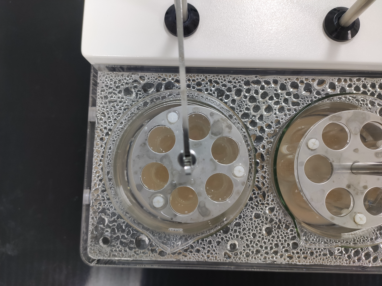
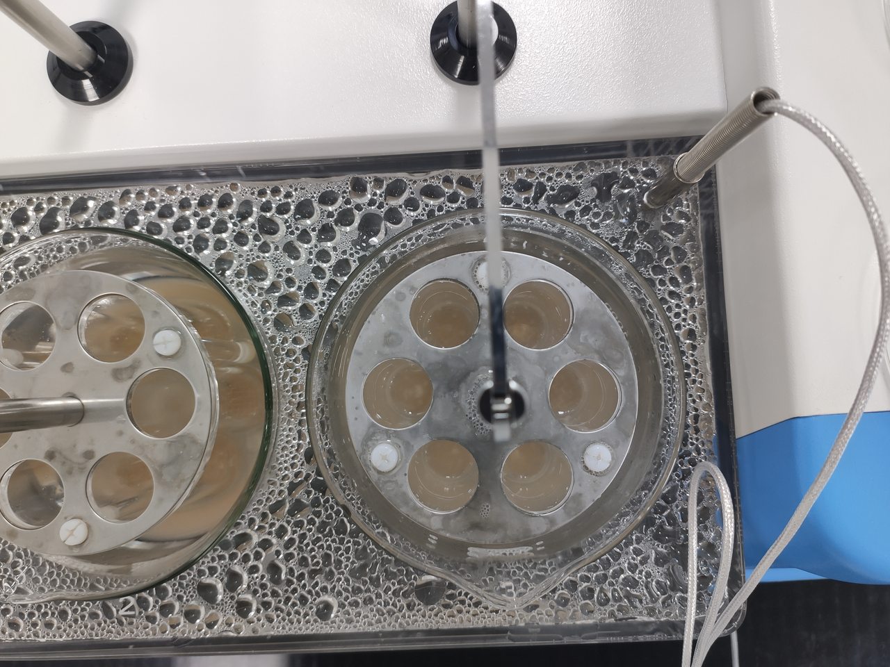
In order to conduct targeted drug stability verification for IBD patients with different colonic pH at different disease activity periods, we formulated artificial intestinal fluid with three pH gradients according to The Chinese Pharmacopoeia. The standardized disintegration time limit of No.2 enteric-soluble hydroxypropyl methylcellulose capsule was examined.
According to the requirements of Chinese Pharmacopoeia, the disintegration verification results of enteric-soluble capsules were obtained by naked eye observation, so we took photos of the experimental process and results to support our naked eye observation results.
After the capsules were filled, we started the experiment of step 1, which was checked in hydrochloric acid solution (9:1000) without baffle for 2 hours. At this time, there was no crack or disintegration of each capsule shell (Fig.24).

Fig.24 A After step 1 is completed.


Fig.25 B Beaker 1 after step 1 C Beaker 2 after step 1 D Beaker 3 after step 1
It can be seen that the capsules in the three beakers did not disintegrate, and the first step was successful.
Then the hanging basket was removed, washed with a small amount of water, and then checked in phosphate buffer solution 1, 2, and 3 in accordance with the above method. All the particles should be disintegrated within 1 hour (Fig.26). If one particle cannot completely disintegrate, another 6 particles should be taken for the retest, which should meet the requirements.

Fig.26 Step 2 in progress.
\Take phosphate buffer 1, 2, and 3 and add them into beakers 1, 2, and 3 respectively. Add 800ml to each beaker and then put them into hanging baskets (Fig.27)

Fig.27 After the completion of Step 2.


Fig.28 A Beaker 1 after step 2 B Beaker 2 after step 2 C Beaker 3 after step 2
In the actual operation, the disintegration time only lasted for 30min, that is, all capsules were found to have disintegrated, indicating that enteric-soluble capsules could disintegrate in the intestinal environment of different pH, and the experiment was successful. (Fig.28 A B C)
This experiment showed that the enteric dissolved capsules we selected could disintegrate and take effect in the intestines of IBD patients with different pH, thus confirming the reliability and stability of the capsules.
[1] Neeloffer Mookherjee K L B D. Human Host Defense Peptide LL-37 Inflammatory Response by the Endogenous Modulation of the TLR-Mediated[J]. The Journal of Immunology, 2021.


