Team:TecCEM/Description

Home

Team

Project

Awards

Human Practices

Parts

Judging form
Project Description
Disrupting Disruptors: A biosensor for the detection of EDCs
Key Concepts: Microplastics, Endocrine Disrupting Chemicals (EDCs), Biosensor, Human Estrogen Receptor Alpha (hER⍺, ESR1), Piezoelectric Sensor.
From all the pollutants, why did we choose Endocrine Disrupting Chemicals?
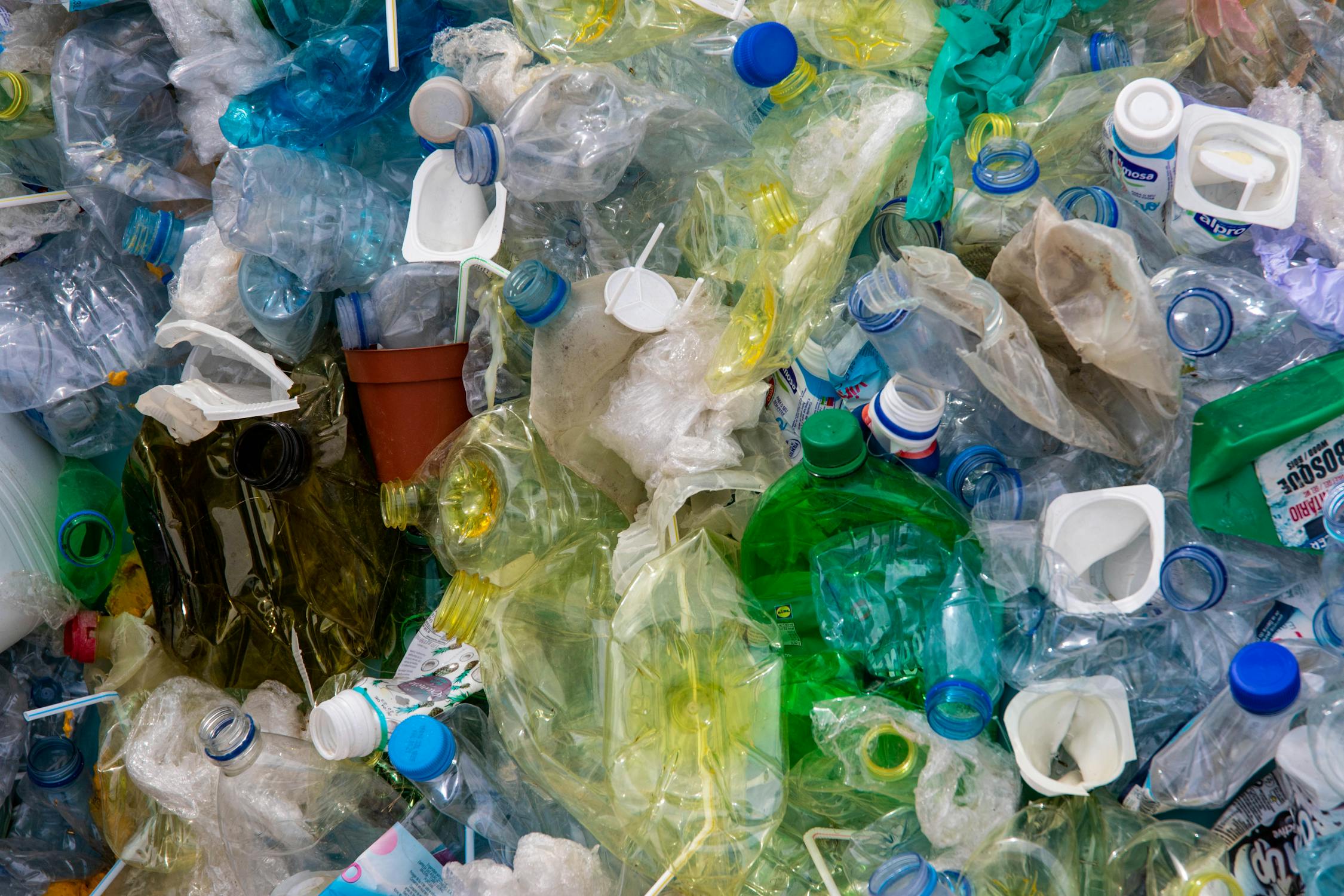

Microplastics represent a serious problem, perhaps they represent one of the biggest dangers of our time. They are generated by the incomplete degradation of materials from cosmetics, bottles, fibers and other plastic related products
[1].
Reference
[1] N. Kalogerakis et al., “Microplastics Generation: Onset of Fragmentation of Polyethylene Films in Marine Environment Mesocosms”, Frontiers in Marine Science, vol 4, bl 84, 2017.
Their size varies a lot as well as its composition; however, it is frequently reported that their size is less than 5 mm and usually come from polymeric materials
[2].
Reference
[2] N. Expósito, J. Rovira, J. Sierra, J. Folch, en M. Schuhmacher, “Microplastics levels, size, morphology and composition in marine water, sediments and sand beaches. Case study of Tarragona coast (western Mediterranean)”, Science of The Total Environment, vol 786, bl 147453, 2021.
More people are becoming aware of the microplastics and and more investigations are focused on trying to solve this problem (including several iGEM teams). Nonetheless, there’s an issue that isn’t being discussed enough: Endocrine Disrupting Chemicals or EDCs.
Are compounds that mimic certain hormones and interfere with body signaling
[3].
Reference
[3] Hormone Health. "Endocrine Disrupting Chemicals EDCs", Health tools for patients and caregivers provided by Endocrine Society, 5-10, 2018.
EDCs are pollutants that come from a variety of sources and are found almost everywhere. They are a whole group of compounds that usually come from the degradation of microplastics and processes related to plastic bottles manufacturing
[4].
Reference
[4] M. Wagner en J. Oehlmann, “Endocrine disruptors in bottled mineral water: total estrogenic burden and migration from plastic bottles”, Environmental Science and Pollution Research, vol 16, no 3, bll 278–286, Mei 2009.
We are as exposed to them as we are to microplastics; it has been reported that in bottled water there’s a substantial amount of EDCs. For some brands of bottled water, some researchers found a concentration of up to 1990 ng/L of EDCs like antimony
[5].
Reference
[5] L. Sax, “Polyethylene terephthalate may yield endocrine disruptors”, Environmental health perspectives, vol 118, no 4, bll 445–448, Apr 2010.
Even though this may not seem like much, we have to remember that these compounds have the potential to bioaccumulate
[6].
Reference
[6] H. Geyer et al., “Bioaccumulation and Occurrence of Endocrine-Disrupting Chemicals (EDCs), Persistent Organic Pollutants (POPs), and Other Organic Compounds in Fish and Other Organisms Including Humans”, vol 2, 2007, bll 1–166.
They have lasting effects on the organism and the ecosystem; for us, they may damage our body or produce troubling diseases such as anomalies in the reproductive systems of both men and women; neurological and learning disorders; problems with fertility; metabolic disorders; obesity and diabetes; cardiovascular problems; and even some types of cancer
[3].
Reference
[3] Hormone Health. "Endocrine Disrupting Chemicals EDCs", Health tools for patients and caregivers provided by Endocrine Society, 5-10, 2018.
In the environment, these chemicals affect animal species that are constantly exposed to them; specially several aquatic species which suffer their toxic effects greatly. The lack of regulations in Mexico and other parts of the world towards these compounds is astounding, given the health and environmental implications they pose. We were mostly concerned that in Mexico, there are no laws that talk about a permitted level of EDCs in drinkable water. Since we have seen that plastic bottles are a primary source of EDCs, it was quite shocking to find out that regulatory agencies aren’t as concerned about this issue as we are. Mainly, we focused on the Norma Oficial Mexicana (NOM or Official Mexican Norm) to check this fact and found nothing on NOM.201-SSA1-2015 “Sanitary specifications of products and services. Water and ice for human consumption”; NOM-127-SSA1-1994 “Environmental health, water for human use and consumption”; and NOM -003-ECOL-1997 “Pollutants in wastewater treatment for public service reuse”.
Currently, there aren’t any methods with the needed quality to detect EDCs in an efficient manner. Most methods are either too expensive or not reliable enough. The most reliable way to detect EDCs is through analytic methods that involve expensive equipment in a laboratory such as mass-based analysis processes like mass spectrometry
[7].
Reference
[7] H.-S. Chang, K.-H. Choo, B. Lee, en S.-J. Choi, “The methods of identification, analysis, and removal of endocrine disrupting compounds (EDCs) in water”, Journal of Hazardous Materials, vol 172, no 1, bll 1–12, Des 2009.
However, it is quite obvious that there’s still a need for a faster, reliable and inexpensive way to measure these compounds in situ. In response to this requirement, some researchers have developed sensors that can detect the presence of EDCs through different methods, such as the production of biosensors. Nevertheless, they have a few limitations which include their lack of standardization, high sensitivity towards interference and difficulty of production
[8],
Reference
[8] A. Bezbaruah en H. Kalita, “Sensors and Biosensors for Endocrine Disrupting Chemicals: State-of-the-Art and Future Trends”, Treatment of Micropollutants in Water and Wastewater, Jan 2010.
not to mention that they may be expensive. The aim of our project? To create a biosensor that has the desirable characteristics we have discussed: a reasonable price so that it can be widely used; reliable results that give the exact concentration of EDCs in a sample of bottled water; specificity towards any EDC; and the ability to detect compounds in a fast way that doesn’t require any other lab equipment.
OUR PROJECT
As stated before, there are a few problems with biosensors. They are very sensitive towards interference and they can get really expensive. There have been researchers that have proposed the use of the Human Estrogen Receptor Alpha (hER⍺, ESR1) and its interaction with other proteins to detect these chemicals and measuring them using a piezoelectric sensor
[9].
Reference
[9] M. Murata, C. Gouda, K. Yano, S. Kuroki, T. Suzutani, en Y. Katayama, “Piezo electric sensor for endocrine-disrupting chemicals using receptor-co-factor interaction”, Anal Sci, vol 19, no 10, bll 1355–1357, Oct 2003.
Other sensors proposed use a Quartz Crystal Microbalance (QCM) with part of the ESR1 protein to detect small quantities of estrogenic substances
[10].
Reference
[10] K. S. Carmon, R. E. Baltus, en L. A. Luck, “A biosensor for estrogenic substances using the quartz crystal microbalance”, Anal Biochem, vol 345, no 2, bll 277–283, Okt 2005.
We got inspired by this research and began the development of our own biosensor which has several characteristics that make it unique.
We designed a biosensor for the detection of Endocrine Disruptive Compounds (EDCs) in samples of bottled water. This biosensor will work through the immobilization of hER⍺ protein receptor on a quartz (the QCM), which is part of a piezoelectric sensor. The protein will capture these chemicals, while the piezoelectric sensor will sense the change of mass through a change of the natural resonance of the quartz. This signal will be received and interpreted by a circuit of our own design and will give us information about the concentration of EDCs on the sample of bottled water. To get an idea of how the protein will be connected to the QCM and how it will work, we recommend checking Figure 1. out.

Figure 1.
Schematic of how our receptor protein ESR1 will be immobilized in a piezoelectric sensor and what is the working mechanism of our sensor. When it is exposed to water without EDCs, it will not catch anything and nothing will be reported. When it is exposed to water with EDCs, the receptor will capture them and the sensor will measure a change of weight. This will help us to quantify the amount of EDCs found in a sample of bottled water. Diagram created with BioRender.com
[11]. [11] BioRender. Diagram created with BioRender. https://biorender.com/Reference
To meet this objective, we modified a version of the Human Estrogen Receptor hER⍺ to capture EDCs and detect them through its their immobilization in a Quartz Crystal Microbalance QCM. We made a prototype designed to measure EDCs concentrations in samples of bottled water (Figure 2).
Figure 2.
First prototype of the hER⍺ EDCs detecting biosensor by iGEM TecCEM.


The design of our protein was based on a PDB crystallized structure of the hER⍺ with a PDB code 2IOG (Protein Data Bank, rcsb.org)
[12]
Reference
[12] K. D. Dykstra et al., “Estrogen receptor ligands. Part 16: 2-Aryl indoles as highly subtype selective ligands for ERα”, Bioorganic & Medicinal Chemistry Letters, vol 17, no 8, bll 2322–2328, 2007.
This protein is a natural occurring receptor found in human cells that acts as an enhancer protein which activates after its binding with its ligand. hER⍺’s natural ligand is estradiol and after the molecule binds in its domain, it will undergo structural changes that activate the receptor by the formation of a dimer. It has been established that the most important amino acids for the correct binding of estradiol and hER⍺ are Gly521, His524, Leu525 and Met528
[13]
Reference
[13] K. Ekena, K. E. Weis, J. A. Katzenellenbogen, en B. S. Katzenellenbogen, “Different residues of the human estrogen receptor are involved in the recognition of structurally diverse estrogens and antiestrogens”, J Biol Chem, vol 272, no 8, bll 5069–5075, Feb 1997.
. After we got the coding sequence of ESR1, we had to decide if it was best to keep only the ligand binding domain or to keep the whole protein. We finally decided to keep the entire protein since we found an immobilization method that required all of it.
We then had to decide what modifications we could make in order to mass produce the protein and get it to the QCM without any structural changes that could induce a different folding. We decided to add a linker sequence of four glycines, 1 serine and 1 cysteine. This last residue would aid us in the immobilization protocol we had planned. We also added a 6X histidine tag and a signal peptide sequence (NSP4) for its purification. This NPS4 sequence comes from part BBa_K3606042. We verified that the protein folded correctly through a modelling tool, then verified that it had a similar structure to the natural protein and later we conducted docking experiments with several EDCs to check if the protein would bind to them. To see what we got, please go to the results section;. we merged our designed Biobrick BBa_K3809010 with BBa_K081005 from the iGEM team from the University of Pavia through the Biobrick Assembly Method. Through this, we were able to create a composite part BBa_K3809012 so that we could express large quantities of our protein.
We completed the design of the hardware and the software for our biosensor. In fact, we created a program that lets users have a real time visualization of the oscillation frequency in order to analyze the output; moreover, we provided a mathematical model to find the exact value of the amount of mass present in the sensor.
Degradation:
For the degradation of EDCs, we found that Laccases had been used for this purpose before. At first we thought that we could design our own Laccase; however, plenty of iGEM Teams had already done this several times. So, we screened different Laccases from different iGEM Teams and got the best results out of one in particular. We decided to use the Laccase from 2012 iGEM Team Bielefeld-Germany BBa_K863010. We established a purification protocol and characterized its activity using a color assay involving methylene blue, malachite green and rose bengal. As control, we used a commercial Laccase from the organism Trametes versicolor. The assay consisted in the observation of the colorants degradation through time using a UV-Vis Spectrophotometer. The protocol we envisioned will allow us to produce this enzyme in large quantities and could have the potential to be used for the degradation of these compounds in a water treatment process. After an interview with two experts working in Water Treatment Companies we found out that if we managed to lower the prices on the treatment of water, it would be more attractive for companies to pursue a better quality of water, so our focus was on the standardization of this process.

Safety Project
In addition to our main project, we wanted to focus on another problem we considered important solving: the overuse of antibiotics in the laboratory. It is no secret that antibiotics are largely used in Genetic Engineering as a selection mechanism. However, this comes at a price, since antibiotic resistant bacteria have been increasing over the last decades. This is no small issue; bacteria could acquire resistance to antibiotics through horizontal gene transfer if we are not careful enough, leading to harder to treat infections and low efficacy medications. Several regulatory agencies advise against the use of antibiotics unless it is absolutely necessary. Such regulatory agencies include the U.S. Food and Drug Administration (FDA), the European Pharmacopoeia 7.0, the Evaluation of Medicinal Products (EMA) and the World Health Organization (WHO)
[14].
Reference
[14] G. Vandermeulen, C. Marie, D. Scherman, en V. Préat, “New generation of plasmid backbones devoid of antibiotic resistance marker for gene therapy trials”, Mol Ther, vol 19, no 11, bll 1942–1949, Aug 2011.
Following this line, we wanted to give a solution that could solve the overuse of antibiotics in our laboratory, so we decided to search for compounds that could be used to select transformed bacteria. Consulting with our PI’s and advisors, we found that if you don’t unfreeze competent bacteria using ice or if it takes you too long to use them, many cells will die due to glycerol exposure. This led us to the hypothesis that we could use glycerol alongside a glycerol degrading enzyme as a new selection marker. We did some research and found that E. coli can actually metabolize glycerol through two different pathways. These pathways could be disrupted with the knockout of some genes involved in both of them. After some research we finally found our inspiration. Lindner, S. et al (2019) had already demonstrated that the knockout of the genes glpK, dhaK, fbp and glpX could lead to the disruption of E. coli by exposure to glycerol. So, they created a new metabolic pathway for glycerol that could potentially reverse the effects of the knockout. We got inspiration from this research and set out to create a new mechanism in which we could use a set of enzymes to create a new marker
[15].
Reference
[15] S. Lindner et al., “A synthetic glycerol assimilation pathway demonstrates biochemical constraints of cellular metabolism”, The FEBS Journal, vol 287, 08 2019.

We designed a novel selection marker which includes the use of glycerol and a modified strain of E. coli. The modified DH5⍺ had the genes glpK, gldA and oxyR substituted by mRFP (as a reporter), Paralcaligenes ureilyticus Dehydratase (PuDHT) and alditol oxidase (aldO) genes. The modified strain can’t metabolize this compound without a selection cassette formed by a plasmid with a katE gene. The proposed design will allow us to reduce the antibiotic selection markers used in laboratory protocols by instead using glycerol; diminishing the risk to human health by an accidental release of antibiotic resistant bacteria.
References:
[1] N. Kalogerakis et al., “Microplastics Generation: Onset of Fragmentation of Polyethylene Films in Marine Environment Mesocosms”, Frontiers in Marine Science, vol 4, bl 84, 2017.
[2] N. Expósito, J. Rovira, J. Sierra, J. Folch, en M. Schuhmacher, “Microplastics levels, size, morphology and composition in marine water, sediments and sand beaches. Case study of Tarragona coast (western Mediterranean)”, Science of The Total Environment, vol 786, bl 147453, 2021.
[3] Hormone Health. "Endocrine Disrupting Chemicals EDCs", Health tools for patients and caregivers provided by Endocrine Society, 5-10, 2018.
[4] M. Wagner en J. Oehlmann, “Endocrine disruptors in bottled mineral water: total estrogenic burden and migration from plastic bottles”, Environmental Science and Pollution Research, vol 16, no 3, bll 278–286, Mei 2009.
[5] L. Sax, “Polyethylene terephthalate may yield endocrine disruptors”, Environmental health perspectives, vol 118, no 4, bll 445–448, Apr 2010.
[6] H. Geyer et al., “Bioaccumulation and Occurrence of Endocrine-Disrupting Chemicals (EDCs), Persistent Organic Pollutants (POPs), and Other Organic Compounds in Fish and Other Organisms Including Humans”, vol 2, 2007, bll 1–166.
[7] H.-S. Chang, K.-H. Choo, B. Lee, en S.-J. Choi, “The methods of identification, analysis, and removal of endocrine disrupting compounds (EDCs) in water”, Journal of Hazardous Materials, vol 172, no 1, bll 1–12, Des 2009.
[8] A. Bezbaruah en H. Kalita, “Sensors and Biosensors for Endocrine Disrupting Chemicals: State-of-the-Art and Future Trends”, Treatment of Micropollutants in Water and Wastewater, Jan 2010.
[9] M. Murata, C. Gouda, K. Yano, S. Kuroki, T. Suzutani, en Y. Katayama, “Piezo electric sensor for endocrine-disrupting chemicals using receptor-co-factor interaction”, Anal Sci, vol 19, no 10, bll 1355–1357, Oct 2003.
[10] K. S. Carmon, R. E. Baltus, en L. A. Luck, “A biosensor for estrogenic substances using the quartz crystal microbalance”, Anal Biochem, vol 345, no 2, bll 277–283, Okt 2005.
[11] BioRender. Diagram created with BioRender. https://biorender.com/
[12] K. D. Dykstra et al., “Estrogen receptor ligands. Part 16: 2-Aryl indoles as highly subtype selective ligands for ERα”, Bioorganic & Medicinal Chemistry Letters, vol 17, no 8, bll 2322–2328, 2007.
[13] K. Ekena, K. E. Weis, J. A. Katzenellenbogen, en B. S. Katzenellenbogen, “Different residues of the human estrogen receptor are involved in the recognition of structurally diverse estrogens and antiestrogens”, J Biol Chem, vol 272, no 8, bll 5069–5075, Feb 1997.
[14] G. Vandermeulen, C. Marie, D. Scherman, en V. Préat, “New generation of plasmid backbones devoid of antibiotic resistance marker for gene therapy trials”, Mol Ther, vol 19, no 11, bll 1942–1949, Aug 2011.
[15] S. Lindner et al., “A synthetic glycerol assimilation pathway demonstrates biochemical constraints of cellular metabolism”, The FEBS Journal, vol 287, 08 2019.
-
Index

Tips
Hover the references