Overview
The goal of this year's iGEM Stockholm team was to create a detection method based on aptamers bound to PCDA vesicles. When the aptamers bind to their target due to mechanical stress the PCDA will change colour indicating that the target is present. Quantification of the intensity of the colour change would be proportional to the concentration of the bacteria present in the skin.
PCDA Polymerisation
Polydiacetylenes (PCDA) are a family of polymers created by the polymerisation of diacetylene creating vesicles or tube structures. (1). Due to their unique chromatic properties, they have frequently been used as a detection method. 10,12-Pentacosadiynoic acid (PCDA) belongs in the family of polydiacetylenes and yields a blue colour in its primary form. Under heat exposure (thermochromism), mechanical stress (mechanochromism) or solvent (solvatochromism), PCDA changes colour from blue to red (2). Our biosensor is based on mechanical stress induction: as mentioned before, it is caused by target binding of the aptamer, which results in a colour change.
Formation of PCDA vesicles
In order to create the PCDA vesicles, we first dissolved the PCDA in chloroform, which we then we evaporated using gentle nitrogen flow. Following this, we dissolved the PCDA in water using sonication. The end product was an emulsion with a white colour. The next step was exposure to UV light to induce polymerisation which in turn results in a colour change from white to blue. This protocol has been used before for the creation of PCDA vesicles (3, 4). The solution was stored at 4 degrees Celcius until further use.

Figure 1: Schematic representation of PCDA-vesicle formation and color change induction.
Conjugation of the PCDA vesicles to the aptamers
The conjugation of the PCDA vesicles to the aptamers is based on the Carbodiimide method. This method is used for the creation of amide bonds between carboxylates and amines by adding dicyclohexylcarbodiimide (DCC). DCC is a crosslinker that creates a reactive intermediate which reacts with nucleophiles like amines resulting in an amide bond (5, 6). In our experiments EDC (1-ethyl-3-(3-dimethylaminopropyl)carbodiimide hydrochloride) was used. EDC is the most popular carbodiimide for conjugating carboxylates with amines and it is used together with NHS (N-hydroxysulfosuccinimide) in order to stabilise the reactive intermediate.
We chose this method since PCDA has carboxyl groups and the aptamer is a small DNA sequence with amine groups. The protocol we followed was based on a paper by Wu et al. (4).

Figure 2: Schematic illustration of the mode of action of our biosensor. PCDA-vesicles conjugated to an aptamer change colour upon target binding.
Bacteria Cultivation
Another important part of the project was the cloning of a biobrick, which was made by assembling two existing parts from the registry: part BBa_K103003 coding for staphylococcal protein A, and part BBa_K1073024 amilGFP. Firstly, the two biobricks had to be expressed and harvested through PCR and gel extraction. After that, the two parts were ligated and assembled into a vector. In order to have a functioning protein, the stop codon that was left between the biobricks had to be removed using site-directed mutagenesis. Finally, the new biobrick was cloned into Top10 competent E. coli cells and expression could be confirmed by observing the colonies. The plasmid was sent for sequencing to verify the plasmid sequence and to validate the part.
SELEX
SELEX or Systematic Evolution of Ligands by Exponential Enrichment is a screening technique based on selecting specific targets from a large pool of random oligonucleotides which could be a mixture of RNA, DNA, single or double-stranded, by an iterative process of separation and amplification. The products of SELEX are DNA or RNA aptamers that mimic the properties of antibodies. They can further bind to a big variety of molecules like proteins, peptides, drugs, organic molecules or even metal ions (7).
The reason why we used aptamers as our detection method this year, is that the development of aptamers is cheaper and more flexible compared to antibodies. Moreover, aptamers are easier to manipulate biochemically. For example, it is simple to add functional groups to the aptamers; in our case we conjugated them to PCDA.
The main principle of making aptamers remains the same no matter the target of interest. The random pool of DNA is incubated with the target. After the incubation, the binding complex is separated from the unbound sequences and then eluted in order to have the DNA sequences only. Sequential positive and negative selection steps allow retrieving the aptamer sequences with the highest sensitivity and selectivity for the target of interest.
Then, an error-prone PCR amplifies the selected aptamers and introduces point mutations in some of the sequences, resulting in a library of variants. Ultimately, we select the sense single DNA strands from the PCR product to build a new library, which will be used in the next SELEX round.
This concludes the first round of SELEX. In order to obtain aptamers with high affinity for the target, multiple rounds of SELEX need to be performed (8).
Cell-SELEX
The first part of the experiment was to create an aptamer that can detect Cutibacterium acnes, a bacteria linked to acne but at the same time belongs in the natural flora of the skin (9). In order to do that, we used two types of C. acnes. More specifically, CCUG 1794 T Cutibacterium acnes (Type I) and CCUG 6369 Cutibacterium acnes (Type II) were used for negative and positive control, respectively. At first, we incubated a random pool of DNA with our CCUG 6369 C. acnes cells after measuring the right amounts of cells by OD600 measurement.
Next, we centrifuged the cell-aptamer mixture and removed the supernatant and, as a result, we had only the cell-aptamer complexes. Then, we eluted the aptamers by heat denaturation and separated them from the cells by centrifugation at 14 000 rpm. Next, we amplified the aptamers with PCR. The end product was a double stranded DNA with the antisense sequence having a biotinylated primer.
This primer was used so we can separate the sense from the antisense sequence using streptavidin beads and NaOH elution. At the end of the first round, we had only a pool of aptamers. In the second round, we used the pool of aptamers from the previous round. We incubated it with the positive and negative bacterial clones; we then followed the same steps as in the first round. The protocol we used was optimised from the protocol from Sefah et al (8) and Simaeys et al (10).

Figure 4: Representation of a bacterial cell membrane with multiple different surface targets, in the form of proteins, lipids or similar.
Bacteria Cultivation
Cutibacterium acnes, formerly known as Proprionibacterium acnes, is a rod-shaped gram-positive commensal skin bacterial species (11).
C. acnes sub-types I and II, used in the cell SELEX experiments, were cultured using Brain Heart Infusion media or Blood Agar.
All C. acnes subtypes, including the aforementioned, are known to be facultatively anaerobic (11). We had to adapt the standard bacteria culture to this metabolic demand. In order to reduce the oxygen in contact with the liquid culture, we used long glass tubes filled with media almost to the top and closed them with sealing lids.
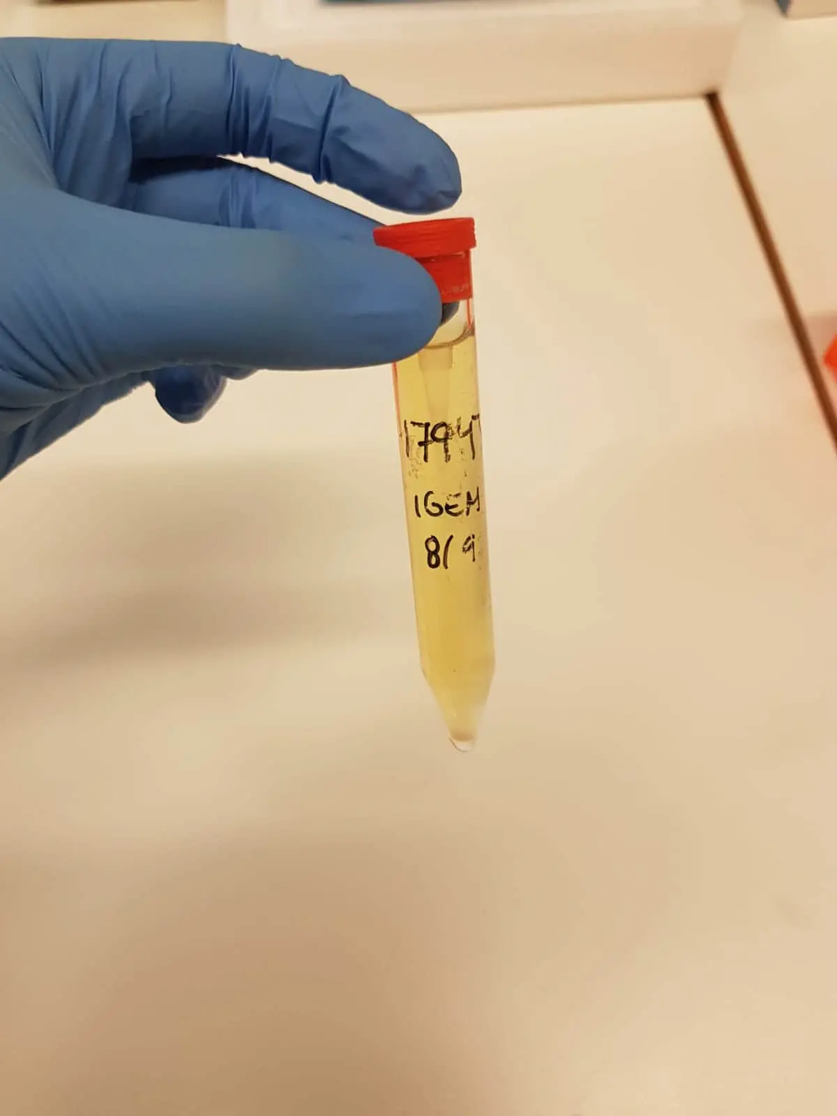
Figure 5: C. acnes cultured anaerobically in Brain Heart Infusion media.
When C. acnes was cultured in plates, we came up with the following strategy: the Blood Agar plates were piled inside of an anaerobic jar. Then, we placed a lit candle on top of the pile of plates and closed the jar with a sealing lid. The candle would light until the oxygen was consumed. This way, we ensured the anaerobic conditions required by the bacteria were maintained inside the jar.

Figure 6: C. acnes culture on a Blood agar plate.
This footage shows how we achieved anaerobic conditions by consuming the oxygen inside the sealed jar.
LTA-SELEX
The second step of our experiment was to create an aptamer that is specific for Lipoteichoic acid (LTA), which is a molecule that exists in the membrane of all gram-positive bacteria (12). Using the carbodiimide method, we conjugated LTA from Staphylococcus aureus to carboxyl-activated sepharose beads. The beads were used as a means of LTA immobilisation and to facilitate the target-aptamer complex separation during the SELEX cycle.
In order to obtain our LTA aptamer, we used the method described earlier, but here we replaced the cells with our LTA-beads as positive control. For the negative control, we used bovine serum albumin (BSA).

Figure 7: Illustration of the cell wall of gram positive bacteria.
Protein-A Aptamer
For our proof of concept, we set out to test a published protein A aptamer. Protein A is a protein that exists in the membrane of S. aureus and an aptamer targeting this specific protein already exists (13). However, when we incubated this aptamer conjugated to PCDA with protein A, we could not observe any colour change. For this reason, we aimed to characterise protein A aptamer binding affinity to our target. To this end, we used HPLC with a protein A column and protein A-bound beads. Unfortunately, both of these methods showed poor target affinity of the aptamer so we tried another approach. We coated wells with protein A and we incubated it with protein A aptamer attached to a fluorophore. From this binding affinity assay, we concluded that our protein A aptamer is not capable of binding protein A efficiently.
BioBrick Engineering
Another important part of the project was building a new BioBrick: BBa_K4071000. The idea was to create a fusion protein consisting of protein A labelled with a reporter protein in order to be visible with the naked eye. Thus, we decided to assemble two pre-existing parts from the registry: part BBa_K1073024, coding for the yellow chromoprotein amilGFP and part BBa_K103003, coding for the Staphylococcal protein A.

Figure 8: Vector map of pSB1C3 containing the amilGFP BioBrick (BBa_K1073024). Retrieved from the iGEM Registry of Standard Biological Parts.

Figure 9: Vector map containing the functional parts of the amilGFP BioBrick (BBa_K1073024).

Figure 10: Vector map of pSB1C3 containing the protein A BioBrick (BBa_K103003). Retrieved from the iGEM Registry of Standard Biological Parts.

Figure 11: Vector map containing the functional parts of the protein A BioBrick (BBa_K103003).
Our cloning strategy was designed based on the BioBrick RFC10 assembly standard:
We decided our BioBrick will first have the amilGFP part, since it contains a promoter and an RBS sequence, and then the protein A construct.
Based on this, we planned to:
- Isolate the protein A CDS
- Linearise the pSB1C3 vector containing amilGFP
- Ligate protein A part into the linearised vector containing amilGFP

Figure 12: Schematic representation of the cloning strategy used to build the amilGFP-proteinA BioBrick (BBa_K4071000). Retrieved from SnapGene.
With these aims, we decided to digest the BioBricks in the following way:
- Protein A BioBrick was digested with XbaI and PstI restriction enzymes:
- This creates sticky ends compatible with SpeI and PstI, respectively.
- Since this part has to go in second position, the BioBrick prefix was removed while the BioBrick suffix was kept.
- AmilGFP plasmid was linearised by digesting with SpeI and PstI restriction enzymes:
- This creates sticky ends compatible with XbaI and PstI, respectively.
- Since this construct has to go in first position, the BioBrick prefix was kept while the BioBrick suffix was removed.
The sticky ends generated in both parts allow directional cloning and regeneration of the BioBrick suffix. Thus, the new BioBrick preserves the standard prefix and suffix needed for RFC10 cloning.
Ultimately, to have a fusion protein, two adjacent STOP codons at the end of the amilGFP CDS had to be removed by using site-directed mutagenesis.

Figure 13: TAA STOP codons (red) at the end of the amilGFP CDS (BBa_K1073024). Retrieved from SnapGene.
Quikchange, a PCR site-directed mutagenesis method, allows the introduction of point mutations in specific locations of interest (14).
The first step we performed was designing two primers that hybridise with the target sequence and, at the same time, contain the modifications we want to incorporate in the template. These primers were then used in a PCR reaction together with a high copy fidelity polymerase, Phusion polymerase in our case. Since the primers have mismatches, the optimal annealing temperature is hard to determine, therefore three different annealing temperatures were used: 55 degrees Celsius, 58 degrees Celsius and 60 degrees Celsius.
Our primers were designed by Kozane.app, a primer design tool for site-directed mutagenesis.
The As in second position in both TAA STOP codons were substituted by Cs in order to create TCA Serine codons. The small size and lack of charge of Serine make the amino acid perfect for this linker region, which should be flexible in order to allow proper folding of both amilGFP and protein A.

Figure 14: Quikchange Forward and Reverse primers are complementary to the target sequence at the end of the amilGFP CDS (BBa_K1073024), but include the desired point mutations (mismatches) that would be incorporated by PCR. Retrieved from SnapGene.
After the PCR, the amplified product consisted of two different populations of DNA molecules: DNA strands which had incorporated the mutation and parental not-mutated strands.
The next step was digesting with DpnI, an endonuclease that recognises methylated and hemimethylated DNA. Since most E. coli strains express the DNA adenine methyltransferase enzyme (15), the parental DNA would be methylated. Therefore, the parental DNA (original template) was susceptible to being digested by DnpI. The PCR product, however, would be unmethylated and would not be digested.

Figure 15: Overview of the Quikchange site-directed mutagenesis method. Image retrieved from Agilent (14).
The last step was transforming TOP10 E. coli competent cells with the mutated DNA molecules, so the bacteria can perform nick repair.
Plasmids extracted from these bacteria would be the newly engineered BioBrick (BBa_K4071000), coding for protein A labelled with the reporter chromoprotein amilGFP.

Figure 16: Vector map of pSB1C3 containing the final version of the amilGFP-protein A BioBrick (BBa_K4071000).
References
Reppy M, Pindzola B. Biosensing with polydiacetylene materials: structures, optical properties and applications. Chemical Communications. 2007 ; (42) : 4317.
Lebegue E, Farre C, Jose C, Saulnier J, Lagarde F, Chevalier Y et al. Responsive Polydiacetylene Vesicles for Biosensing Microorganisms. Sensors. 2018 ; 18 (2) : 599.
Wen JT, Bohorquez K, Tsutsui H. Polydiacetylene-coated polyvinylidene fluoride strip aptasensor for colorimetric detection of zinc (II). Sensors and Actuators B: Chemical. 2016 Sep 1 ; 232 : 313 - 7.
Wu W, Zhang J, Zheng M, Zhong Y, Yang J, Zhao Y, Wu W, Ye W, Wen J, Wang Q, Lu J. An aptamer-based biosensor for colorimetric detection of Escherichia coli O157: H7. PloS one. 2012 Nov 7 ; 7 (11) : e48999.
Shah TR, Misra A. Proteomics Challenges in Delivery of Therapeutic Genomics and Proteomics.
Hermanson GT. Bioconjugate techniques. Academic press; 2013 Jul 25.
Chai C, Xie Z, Grotewold E. SELEX (Systematic Evolution of Ligands by EXponential Enrichment), as a powerful tool for deciphering the protein–DNA interaction space. InPlant Transcription Factors 2011 (pp. 249 - 258). Humana Press.
Sefah K, Shangguan D, Xiong X, O'donoghue MB, Tan W. Development of DNA aptamers using Cell-SELEX. Nature protocols. 2010 Jun ; 5 (6) : 1169 - 85.
Spittaels KJ, Ongena R, Zouboulis CC, Crabbe A, Coenye T. Cutibacterium acnes phylotype I and II strains interact differently with human skin cells. Frontiers in Cellular and Infection Microbiology. 2020 Nov 16 ; 10 : 712.
Van Simaeys D, Lopez-Colon D, Sefah K, Sutphen R, Jimenez E, Tan W. Study of the molecular recognition of aptamers selected through ovarian cancer cell-SELEX. PloS one. 2010 Nov 1 ; 5 (11) : e13770.
Elston MJ, Dupaix JP, Opanova MI, Atkinson RE. Cutibacterium acnes (formerly Proprionibacterium acnes) and Shoulder Surgery. Hawaii J Health Soc Welf. 2019 ; 78 (11 Suppl 2) : 3 - 5.
Poxton IR. Teichoic acids, lipoteichoic acids and other secondary cell wall and membrane polysaccharides of gram-positive bacteria. InMolecular Medical Microbiology 2015 Jan 1 (pp. 91 - 103). Academic Press.
Stoltenburg R, Krafcikova P, Viglasky V, Strehlitz B. G-quadruplex aptamer targeting Protein A and its capability to detect Staphylococcus aureus demonstrated by ELONA. Scientific reports. 2016 Sep 21 ; 6 (1) : 1 - 2.
Agilent. QuikChange Site-Directed Mutagenesis Kit Instruction Manual. Available from: https://www.agilent.com/cs/library/usermanuals/public/200518.pdf [Accessed 14 Oct 2021].
New England Biolabs. Dam and Dcm Methylases of E. coli. Available from https://international.neb.com/tools-and-resources/usage-guidelines/dam-and-dcm-methylases-of-e-coli [Accessed 14 Oct 2021].