Team:MADRID UCM/Encapsulation
![]()

Encapsulation
Encapsulation of biological entities is now being widely used in many sectors, such as in medical research, food or biotechnology industry. We have used the Encapsulation as a method to introduce our cells within a material in order to create an environment where cell survival is enhanced and metabolism can be easily routed towards product synthesis.
In this page you will find all the information about the differen bio-hybrid materials we have studied to obtain a cyanobacterial-powered solid photobiocatalysts.
What is cell encapsulation?
Encapsulation is a process that consists of the confinement of an entity to a limited region of space, defined by the boundaries of an encapsulation material. Within biocatalyst synthesis it is considered as one immobilization technique, where a biological entity is confined inside a material. Encapsulation is one of the easiest ways for the creation of whole-cell biocatalysts. An emerging field that aims to achieve the same advantages that enzyme immobilization achieved for the creation of biocatalysts and biosensors. Then, cell encapsulation consists in the creation of a biohybrid material, where cells are entrapped within.
This way, cell encapsulation can be performed for multiple purposes… to protect the cells inside the material, to control the secretion of a product synthesized by the cells or to simply generate a biohybrid material with attractive properties. If you have been reading other parts of the web, you will already know what we are going to encapsulate....CYANOBACTERIA!
Advantages of encapsulation
Encapsulation is a versatile technique that may offer important advantages...
1. It is a good and reliable method to enhance the catalytic properties of biological materials. Since most biological catalysts are small enough to perform in single-phase homogeneous systems, its recovery is challenging. Encapsulation eases its recovery and reutilization greatly reducing the amount of required catalyst.
2. Biological entities are more fragile than traditional industrial catalysts. Encapsulation can provide protection against environmental threats for them. As an example, encapsulation materials can filter UV or other radiations from the environment, can provide protection against contaminants or heavy metals present in the media (which are size-excluded and cannot reach the encapsulated cells). In addition, encapsulation provides a scaffold which protects the cells from external mechanical stress.
3. Encapsulation can greatly stabilize the biological material, extending its life-span. In the case of whole cells, metabolism can be altered, where in some cases higher productivity of extracellular metabolites has been demonstrated. Since cells cannot divide, the surplus of available energy and materials is channelized towards product formation. In addition, cell survival can also be extended, where drying periods, harsh pH or temperature changes can be easily tolerated by the encapsulated cells.
4. Eventually, when living cells are encapsulated, the risk of being attacked by other organisms is significantly reduced. In a similar fashion, when using engineered microorganisms, encapsulation can also significantly reduce the release risk, acting as a passive biocontainment measure.
How the ideal photobiocatalyst looks like?
Encapsulated whole-cell biocatalysts can be produced in many different ways, depending on the selected materials for encapsulation and synthesis procedures. However, in spite of the wide range of available options, the development of a whole cell photobiocatalysts is a challenging process, since those biohybrid materials require the simultaneous combination of multiple features.
- Biocompatibility. The encapsulation material must be compatible with the cells to be encapsulated. In other words,it must be possible that the cells perform their biological functions inside (with the exception of reproduction, given the encapsulation system to which they are subjected) and toxic or harmful effects due to the material are limited.
- High diffusivity. A low diffusional limit for encapsulation materials is one of the key requisites for the design of any catalyst. The cell must be able to adequately acquire the necessary nutrients from the liquid media surrounding catalyst particles. In addition, the product must be able to be released through the encapsulation envelope. This requirement shall be pursued through pore size modification. In general terms, the larger the pore size, the lower the diffusional limit. However, small pores could also be required to efficiently encapsulate the cells within the materials, avoiding cell leakages.
- Transparency. Besides the capacity of efficiently allowing the cells to exchange materials with their surrounding environment, in a whole-cell photobiocatalyst cells will also require light. Then, encapsulation materials must be transparent or translucent, in order to provide the cells with the energy required to perform their functions.
- Good mechanical properties. In order to easily separate the biocatalyst from the media so that the catalytic activity of the cells is retained and can be reused. The material must be robust enough to keep on with mechanical stress during its manipulation. Likewise, insoluble encapsulation materials should be used.
- Environmentally friendly. There are numerous advantages of photobiocatalytic materials, notably that they produce no waste during catalysis, reduce carbon emissions and can be recycled. However it is crucial to consider materials which can be easily obtained, are abundant and can be easily recycled. Even the utilization of biodegradable materials must be considered, when after photobiocatalyst depletion it can be processed as organic waste.
Our Encapsulation Systems
Keeping in mind all of the above features that an ideal biocatalysts will require, we performed a deep research in order to select our encapsulation materials. After this, we proposed a set of biohybrid materials that could become good photo biocatalysts. Then, with the advice of the Biohybrid Materials group of the Materials Science Institute of Madrid (ICMM-CSIC), we designed the synthesis protocols for each material proposed.
Explore Our Materials
Alginate-Sepiolite Beads

The beads are small porous spheres in which the cells can lodge. They are created with alginate, a biopolymer that, in the presence of calcium, creates a structure known as an 'egg crate' where cyanobacteria can be internalised, resulting in a material with reduced porosity, distributed in cavities in the range of 50-200 nm. Additionally, in order to increase biocompatibility, sepiolite, a clay, is added.
The alginate-sepiolite beads are spheres of approximately 0.2 cm diameter inside which the cells are housed. Sepiolite is included in the material in order to increase biocompatibility.
This material has been created by producing mixtures of different proportions of the two reagents and then, using a tipped syringe, adding the mixture dropwise to a calcium chloride solution, instantly creating hardened alginate-sepiolite beads.
The cell biomass is added prior to depositing the mixture in the calcium chloride, and always under the best possible sterile conditions.
After a few minutes, they are removed from the calcium chloride and placed in BG-11 culture medium.
Our research
The beads meet the expectations we had at the beginning of the research, they are resistant, they do not fall apart in the environment and the cells survive in them.
Problems are observed in the diffusion of the product to the outside, probably due to the pore size.
To preserve cell viability, it is necessary to keep them in a sterile container with natural light and room temperature.
Evaluation as Photobiocatalyst: Pros & Cons
Biocompatible with the cells ✅
Transparent ✅
Good porosity and low diffusional limitations ❌
Good mechanical properties, resistance and stability ✅
Easy Separation ✅
Biodegradable ✅
Through the microscope

Explore more materials
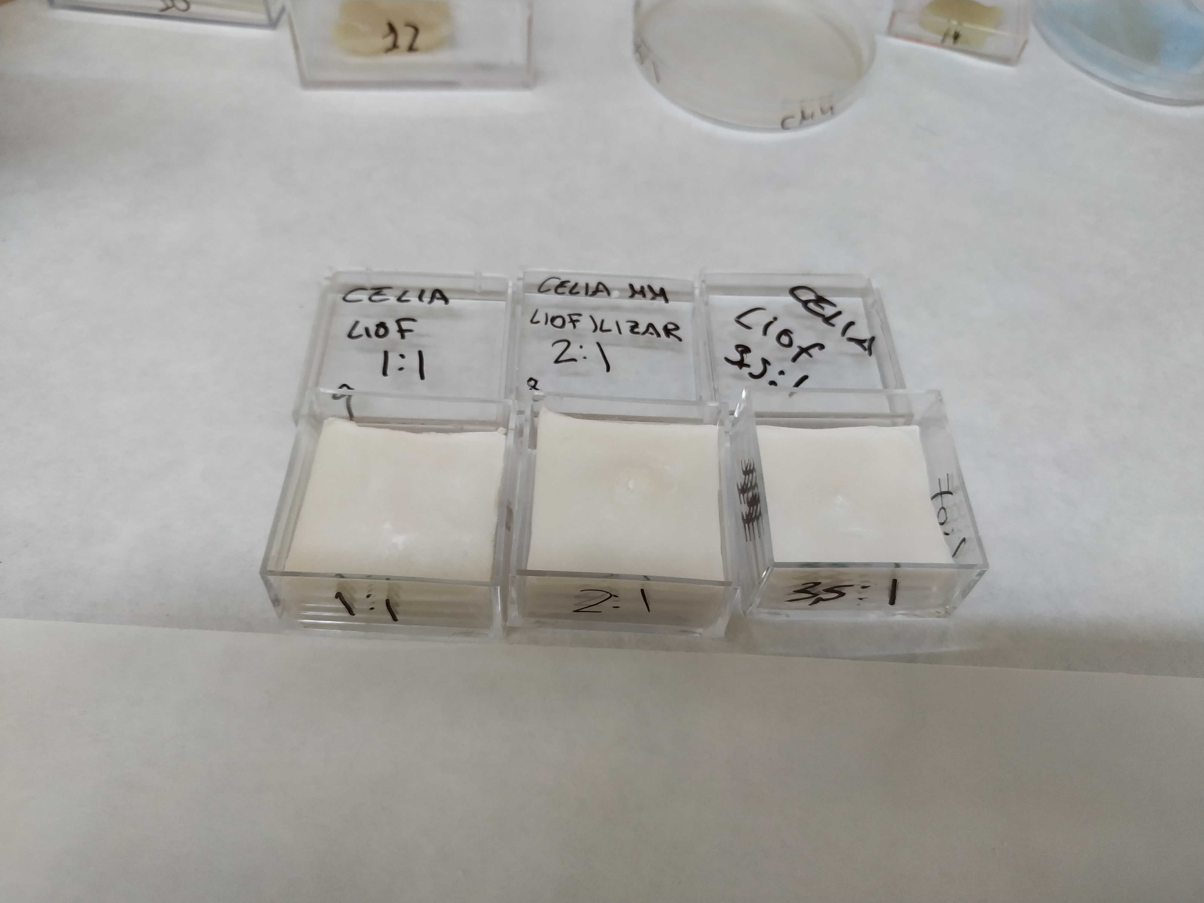
Chitosan-Sepiolite Thin Films

Thin films are, as the name suggests, thin strips of (ideally resistant) material in which the cyanobacteria are encapsulated. Thin films are an alternative to beads, designed to have a larger amount of cyanobacteria in a smaller amount of material, as this type of material takes up very little space, being very thin and resistant 'strips'. Moreover, these thin films can be created in any size desired, and are designed with the idea in mind that after this encapsulation, the aim is to introduce the materials into a photobioreactor. Thus, with thin films, a large number of encapsulated cells can be placed in a minimum size. In addition, it is a very transparent and resistant material. Chitosan, a biopolymer, is used for its formation, but unlike alginate, it will not dissolve in water when creating the thin films; with sepiolite to increase biocompatibility, as we did with beads.
Solutions of chitosan and sepiolite were used as a starting point, and the two were then mixed in different proportions until a homogeneous mixture was achieved. To this mixture, the previously centrifuged cells were added, as in all the materials, and then, using a syringe, they were poured onto a flat surface (Petri dishes or similar were used). Between 24 and 48 hours, a thin, resistant and malleable layer is obtained, which is placed in BG-11 for preservation.
Our research
Although thin films were not initially a material in which we had a lot of confidence, good results have been obtained from working with them.
It is remarkable for its resistance in spite of its small thickness, which also confers them a low diffusional limitation in spite of its reduced porosity.
Furthermore, it is important to point out that the higher the m/v percentage of chitosan, the better the mechanical properties and stability of the material.
Evaluation as Photobiocatalyst: Pros & Cons
Biocompatible with the cells 🟧
Transparent ✅
Good porosity and low diffusional limitations ❌
Good mechanical properties, resistance and stability ✅
Easy Separation ✅
Biodegradable ✅
Through the microscope

Explore more materials
Biopolymer-Sepiolite Solid Foams
In order to improve cell strength and cell durability over time, it is thought that foams can improve certain properties of thin films, such as pore size. This type of material has been created following a freeze-drying process, using materials such as chitosan and sepiolite, as in thin films. The foams, unlike the rest, are macroporous materials.
They could be easily handled, resistant and moderately translucent materials, which could be moved almost anywhere. Their main problems are its limited transparency and their resistance to water.
These materials are produced by freeze-drying a suspension of biopolymer and sepiolite.
Therefore, after producing the foams, the cyanobacteria are introduced inside by pressure, using a syringe.Interaction of the cells with the sepiolite clay may be sufficient to keep them entrapped within the material.
Our research
Despite the fact that at first, foams were one of the materials that we thought would have the best properties for surface immobilization of cells, their lack of consistency and their poor resistance in water or BG-11, makes us reconsider other ways of creating this material in order to use it as a biocatalyst.
However its highly porous structure is still being a promising alternative to act as casing for other materials. To achieve this goal, other polymers should be used in order to increase its hydrophobicity.
Evaluation as Photobiocatalyst: Pros & Cons
Biocompatible with the cells ✅
Transparent 🟧
Good porosity and low diffusional limitations ✅
Good mechanical properties, resistance and stability ❌
Easy Separation ✅
Biodegradable ✅
Through the microscope

Explore more materials
Yolk-Shell Microencapsulation

Yolk-shells are microscopic materials where encapsulation is performed at the single cell level.
They allow the creation of a silica 'protective shield' around the cells, which, with the addition of a protein called protamine, creates a space between the cell and the shield, allowing for greater biocompatibility and less toxicity to the cells as they are not closely related to the silica.
This is possible because the cyanobacteria internalise the protamine, 'eating' it little by little, leaving a gap in the end.
This will allow us to have the cells encapsulated easily, in a medium as if it were a normal culture. In addition, they can be used in the same way as if they were not encapsulated, allowing us to improve some encapsulation techniques such as thin films or silica gels, by being able to introduce cells that are already protected.
To create this material, colloidal self-assembly of silica nanoparticles is performed. Protamine is used to coat the cells during colloidal self-assembly, and once the cyanobacteria has internalised it, it will leave a gap between the silica coating and the cell.
The protocol involves successive centrifugations and resuspensions of the culture in PBS buffer, the addition of protamine sulphate, and colloidal silica nanoparticles (LUDOX-TMA ® )
Our research
This material, not only has good results in its own right, but also we are sure that mixed with other materials, such as gels or thin films could significantly improve its performance.
The observable results are quite good. The material retains the normal green color of the cells and even produces gas bubbles when illuminated.
Given their size, it is not visible to the naked eye whether the cells have been successfully coated. However, after less than one hour, they settle to the bottom of the container. This behaviour is not usual in normal cells and is the first confirmation of a successful encapsulation that can be further confirmed by electron microscopy examination.
We have also assessed the qualitative long term cell viability considering the relative chlorophyll content. They have been found to lose their colour in PBS in the matter of a week, while they retained their colour and continued producing gas bubbles, proofs that the material can successfully keep cells alive for long periods of time.
Evaluation as Photobiocatalyst: Pros & Cons
Biocompatible with the cells ✅
Transparent ✅
Good porosity and low diffusional limitations ✅
Good mechanical properties, resistance and stability ✅
Easy Separation 🟧
Biodegradable 🟧
Through the microscope

Explore more materials
Biocompatible Silica Gels

The silica gels constitute a highly porous three-dimensional SiO2 network, with a mesoporous distribution of porosity, with a high number of micropores and mesopores of a size close to 20 nm. The cells are "encapsulated" in the material as they are embedded in the matrix, establishing almost direct contact with it.
This type of material can be created in two stages of synthesis. First, the creation of a silica precursor by ion exchange of sodium silicate solutions. Second the cells are dispersed into it and the creation of the gel by addition of a basic pH-modifying reagent. Initial ion exchange step performed to avoid the toxicity that an excess sodium ion may pose to the cells.
They are obtained by the ion exchange of sodium silicate solution, the gel precursor. Sodium ions are removed in order to increase biocompatibility.
Subsequently, silica nanoparticles and potassium hydroxide are added,adjusting the pH to be the ideal for the cells.Then, biomass is added and evenly distributed.
Finally, by keeping moderate heating (30-38ºC) and mild agitation for even biomass distribution the gel is formed and will become solid.
Extreme drying of the gel should be avoided to guarantee adequate cell viability.
Our research
Despite the various problems that have arisen through the process of preparing and fine-tuning protocols, it has proved to be a material with notably better properties than others previously described.
The synthesis protocol is challenging, since small variations in gelification times, amount of pH reagent used and sterility may severely alter the overall viability of the cells within the material.
It is important to note that the best point to have these materials is in an intermediate point between the gel (like gelatine) and crystal-like dry aspect.
*Silica gels with yolk-shell: In order to generate a material with the benefits shown by yolk-shell at a macroscopic scale, we have mixed both materials. The results of this complex yolk-shell silica gel materials are even better than silica gels, where cells are efficiently entrapped and leakage is not observed.
Evaluation as Photobiocatalyst: Pros & Cons
Biocompatible with the cells ✅
Transparent ✅
Good porosity and low diffusional limitations ✅
Good mechanical properties, resistance and stability ✅
Easy Separation ✅
Biodegradable 🟧
Through the microscope

Explore more materials
Developing a photobiocatalyst
At this point, you may already have an idea of how a photobiocatalyst could look like. But, how can we evaluate the performance of the proposed materials? We need to create a material where cyanobacteria can live, capture carbon dioxide and secrete the product of interest and withstand the conditions found within a cell cultivation setup. Only by studying these aspects can we answer the question of how our materials end up as a biocatalyst?
Characterizing our materials
After fine-tuning the synthesis protocol of each material, the next step is the detailed evaluation of its properties, to do so we have performed multiple experiments.
1. First of all, the catalyst needs to be compatible with cells. So, it is important to always present that one of the most relevant things is cell viability and robustness.
2. Second step is to test diffusivity. How can we do that? Designing an assay where our product of interest or a molecular with similar properties is forced to diffuse through the material to the surrounding media. Studying the time -concentration profiles of the diffusing species will allow us to know which material is the best in terms of diffusivity. This test has been proved with beads, thin films and silica gels.
3. Finally, when all materials have been synthesised, it is very important to physically characterise them. What does this mean? Characterisation helps us to study different properties of materials that provide us with information about them. For this purpose, the different catalysts created have been viewed by SEM and optical microscopy. In addition, nitrogen adsorption isotherms and mercury porosimetry will also be desirable to assess the pore size and distribution within the material.

To know more about the material characterization part, you can visit the Experiments and Results pages. Likewise, if you want to read about the protocol optimization and synthesis procedures, you can download the encapsulation notebook.


Conclusions
As an important part of our project, we have designed and evaluated multiple cell encapsulation protocols to create a photobiocatalytic material. After researching, designing and evaluating multiple materials the most important conclusions can be highlighted as:
- We have been able to manufacture a photobiocatalytic material with enhanced production properties in comparison with free cells.
- From all of the proposed materials, the yolk-shell structures may be the most suitable for direct industrial applications. This material retains all the beneficial properties from free cells, while extending its life span, protecting them from the environment threats, increasing its productivity and allowing its utilization at higher densities than conventional cells. Further research will be required to assess the influence of encapsulation in their metabolic performance.
- Silica gels have also demonstrated interesting properties, however the accuracy required for its successful synthesis without compromising cell viability is still a challenge to solve.
- We have provided the iGEM community with a starting point for the development of biohybrid materials based on the whole-cell encapsulation concept. While the utilization of these materials as catalysts for biomanufacturing requires a challenging set of features, all of the proposed materials could be useful for other applications such as biosensors.
Encapsulation of living entities
M. Flickinger, J. Schottel, D. Bond, A. Aksan and L. Scriven, "Painting and Printing Living Bacteria: Engineering Nanoporous Biocatalytic Coatings to Preserve Microbial Viability and Intensify Reactivity", Biotechnology Progress, vol. 23, no. 1, pp. 2-17, 2007. Available: 10.1021/bp060347r
J. Rooke, A. Léonard, C. Meunier and B. Su, "Designing Photobioreactors based on Living Cells Immobilized in Silica Gel for Carbon Dioxide Mitigation", ChemSusChem, vol. 4, no. 9, pp. 1249-1257, 2011. Available: 10.1002/cssc.201000442
S. Vasilieva, E. Lobakova, A. Lukyanov and A. Solovchenko, "Immobilized microalgae in biotechnology", Moscow University Biological Sciences Bulletin, vol. 71, no. 3, pp. 170-176, 2016. Available: 10.3103/s0096392516030135
Nanocomposites and general materials:
E. Ruiz-Hitzky, M. Darder, A. Alcântara, B. Wicklein and P. Aranda, "Recent Advances on Fibrous Clay-Based Nanocomposites", Organic-Inorganic Hybrid Nanomaterials, pp. 39-86, 2014. Available: 10.1007/12_2014_283
M. Darder, P. Aranda and E. Ruiz-Hitzky, "Bionanocomposites: A New Concept of Ecological, Bioinspired, and Functional Hybrid Materials", Advanced Materials, vol. 19, no. 10, pp. 1309-1319, 2007. Available: 10.1002/adma.200602328
C. Meunier, J. Rooke, A. Léonard, H. Xie and B. Su, "Living hybrid materials capable of energy conversion and CO2 assimilation", Chemical Communications, vol. 46, no. 22, p. 3843, 2010. Available: 10.1039/c001799j
D. Killeen, M. Frydrych and B. Chen, "Porous poly(vinyl alcohol)/sepiolite bone scaffolds: Preparation, structure and mechanical properties", Materials Science and Engineering: C, vol. 32, no. 4, pp. 749-757, 2012. Available: 10.1016/j.msec.2012.01.019
P. Johnson et al., "Spray-Dried Multiscale Nano-biocomposites Containing Living Cells", ACS Nano, vol. 9, no. 7, pp. 6961-6977, 2015. Available: 10.1021/acsnano.5b01139
Silica based materials
M. Perullini, M. Calcabrini, M. Jobbágy and S. Bilmes, "Alginate/porous silica matrices for the encapsulation of living organisms: tunable properties for biosensors, modular bioreactors, and bioremediation devices", Open Material Sciences, vol. 2, no. 1, 2015. Available: 10.1515/mesbi-2015-0003
J. Rooke, A. Léonard, H. Sarmento, C. Meunier, J. Descy and B. Su, "Novel photosynthetic CO2bioconvertor based on green algae entrapped in low-sodium silica gels", J. Mater. Chem., vol. 21, no. 4, pp. 951-959, 2011. Available: 10.1039/c0jm02712j
D. Dickson and R. Ely, "Silica sol-gel encapsulation of cyanobacteria: lessons for academic and applied research", Applied Microbiology and Biotechnology, vol. 97, no. 5, pp. 1809-1819, 2013. Available: 10.1007/s00253-012-4686-8
M. Darder et al., "Algae–silica systems as functional hybrid materials", J. Mater. Chem., vol. 20, no. 42, pp. 9362-9369, 2010. Available: 10.1039/b913269d
L. Wang et al., "Single-cell yolk-shell nanoencapsulation for long-term viability with size-dependent permeability and molecular recognition", National Science Review, vol. 8, no. 4, 2020. Available: 10.1093/nsr/nwaa097