Despite the fact that cancer diseases and resulting possible treatment options are being intensively studied, the illness remains one of the leading causes of death worldwide. Tumors are unfortunately very diverse, and therapies used for many cancers, such as chemotherapy, are often aggressive and cause severe side effects. Thus, new treatment options are needed that specifically target solely tumor tissue without damaging the rest of the body. This would greatly benefit healing prospects, reduce side effects to a minimum and help patients to regain life quality.
Since bacteria can be modified easily, they are conceived as potential cancer therapeutics. Attenuated Salmonella spp (S.) are among the intensively studied options for drug delivery vehicles and they show high safety and efficacy in murine models.1
Derived from this approach, bacterial minicells are of special interest to serve as a basis for therapeutics cargo. They can be equipped with a variety of properties and show no toxicity or immunogenicity in different model species. Thus, they are a considerable option in personalized cancer therapies.2
To date, minicells are mainly used in approaches in which they must be endocytosed or are delivering drugs to the microenvironment of tumors,3 i.e. they have to enter the cell or the drug is diluted in the microenvironment.
We aimed to combine independent former research findings to develop a minicell-based system able to deliver a peptide agent directly and specifically into tumor cells without the need of being phagocytosed.
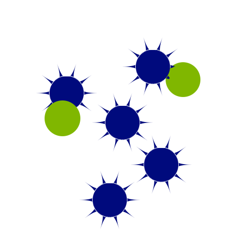
We want to utilize S. Typhimurium-derived minicells as a shuttle – or taxi system – to fight cancer. We produce minicells by inducing asymmetric cell division in S. Typhimurium cells. This is achieved by deletion of minD, a gene encoding for a part of the Min system resulting in the constriction of small, round and nucleoid-free cells.4 Minicells are about 0.4–0.5 µm in size.5 It has been demonstrated that they can be packed with 1–10 million drug molecules.6 Satisfactorily, since minicells lack the chromosome they are not able to divide, thus ruling out the risk of pathogenic infection in clinical application. To gain a minicell sample without parental cells while maintaining high yields of minicells, we refined an existing minicell purification protocol.7 Furthermore, bacterial minicells still harbor components necessary for transcription and translation as well as metabolic machinery. Thus, minicells can be equipped with plasmids from which different genes can be expressed. As a result, these small cells are able to survive and express genes for several hours. This should allow the minicells to survive until they reach the designated tumor tissue and enable them to fulfill a given task: e.g. produce a peptide effective against cancer cells at the target site.
Since minicells are small and blood vessels within tumor tissue are leaky, minicells have the tendency to accumulate in the tumor microenvironment which represents a passive targeting mechanism.2 To also allow active targeting of cancer cells, we aimed to display a lectin domain on the surface of the minicells, which should boost the attachment of minicells to the cancer cells. For this, we plan to clone the recombinant N-terminal domain of the lectin BC2L-C of Burkholderia cenocepacia (rBC2LCN). This lectin domain was shown to bind to surface glycans of several tumor cells, e.g. MCF7 breast cancer cells.8 If it is possible in the future to display recombinant lectins on the surface of minicells, this could be a promising approach for active targeting of cancer cells and would represent an advantage over commonly used antibodies. For the display of the lectin we designed two different approaches, which have to be evaluated in the future. In one of them, the lectin is inserted between the signal sequence of the AIDA-I autotransporter and the autotransporter itself.9 In the other approach, the lectin is inserted between the signal sequence of the adhesin component FimH and the secretory part of it.10 Both systems have been shown to allow for display of exogenous proteins on the bacterial surface, for the AIDA-I autotransporter even on minicells.
As an anti-cancer agent, we wanted to use the synthetic peptide Pep8.11 Pep8 suppresses the growth of MCF-7 cells, as shown previously.11 To translocate it into the cancer cells, the injectisome of the Salmonella pathogenicity island 1 (SPI-1) was planned to be used. In the SPI-1, virulence genes for the invasion of host cells are located.12 In addition to genes encoding components of the injectisome, genes of effectors are also localized in SPI-1, for example the genes encoding SptP and its chaperon SicP. SptP harbors an N-terminal secretion tag (SptP167). It was shown previously that the translational fusion of a protein of interest to this secretion tag allows for translocation of heterologous proteins via the type III secretion system (T3SS) of the SPI-1 injectisome.13 To enable synchronized expression and translocation of our Pep8 only at the site of interest: i.e. tumor tissue, we planned to place the construct under the control of a lactate inducible promoter. It was recently described that the lactate concentration is much higher in the tumor microenvironment compared to blood and healthy tissue, thus enabling our system to produce Pep8 only when the minicells have reached cancerogenic tissue.14 Furthermore, our plasmid-based expression system of Pep8 is not based on a standard applied backbone in lab routine. We deliberately designed, created and evaluated a novel plasmid fitting our needs. Because standard plasmids require constant selection pressure e.g. antibiotics, in order for cells to maintain the backbone this type of selection pressure was not feasible for our approach. Instead, we evaluated the selection system based on the compilation of an essential gene in Salmonella. Our results show that by this, minicells can be stabley equipped with our newly created plasmid and plasmid-based expression is possible.15
List of Sources
Project Description
Development of the Idea

Phase 1: Production and characterization of minicells

Phase 2: Adhesion of minicells to cancer cells

Phase 3: Injection of an anti-cancer peptide into cancer cells
